SciELO - Scientific Electronic Library Online

 
vol.82 número1Achados tricoscópicos na micose fungóide foliculotrópica: a propósito de um casoUm caso de melanose de Becker na fronte: uma apresentação rara índice de autoresíndice de materiabúsqueda de artículos
Home Pagelista alfabética de revistas  

Servicios Personalizados

Revista

Articulo

Indicadores

Links relacionados

  • No hay articulos similaresSimilares en SciELO

Compartir


Portuguese Journal of Dermatology and Venereology

versión impresa ISSN 2795-501Xversión On-line ISSN 2795-5001

Port J Dermatol Venereol. vol.82 no.1 Lisboa mar. 2024  Epub 18-Sep-2023

https://doi.org/10.24875/pjdv.23000051 

DERMATOLOGY IMAGES

A rare case of granulomatous slack skin with scarce multinucleated giant cells

Um raro caso de cútis laxa granulomatosa com escassas células gigantes multinucleadas

Laísa E. de Hollanda1  https://orcid.org/0000-0003-4240-0777

Carolina S. de Oliveira1 

Isabella C. Mendes-Alexandre1 

Rebeca de O. Alves-Melo-Martins1 

Vanessa R. Ferreira1 

Monique F. dos Reis2 

Silvana de A. Damasceno-Ferreira3 

Luciana M. dos Santos1 

1Department of Dermatology, Hospital Universitário Getúlio Vargas

2Department of Pathology, Hospital Universitário Getúlio Vargas

3Department of Pathology, Fundação Hospitalar de Dermatologia Tropical e Venereologia Alfredo da Matta. Manaus, AM, Brasil


A previously healthy 23-year-old woman was observed with erythematous and infiltrated plaques associated with significant skin flaccidity forming a pendulous skin fold in the right armpit and a brown macular pigmentation with mild skin flaccidity in the left armpit (Fig. 1). Lesions were asymptomatic and had a 2-year evolution.

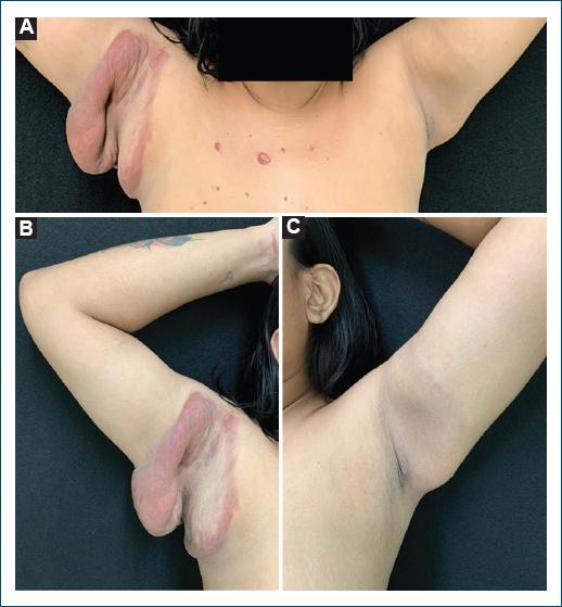
Figure 1 A: skin lesions in both armpits with a 2-year evolution. B: erythematous and infiltrated plaques associated with significant skin flaccidity in the right armpit. C: brown macular pigmentation with mild skin flaccidity in the left armpit. 

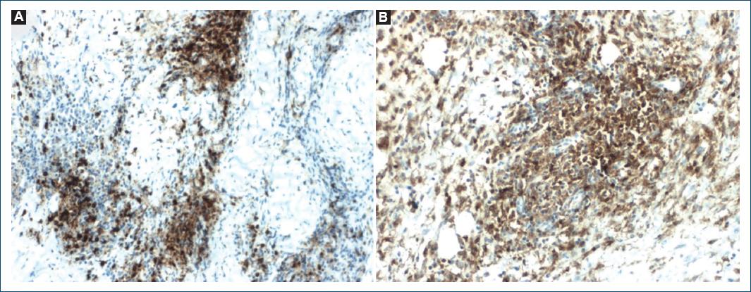
A skin biopsy of the right armpit revealed a dense dermal granulomatous infiltrate of atypical lymphocytes, neutrophils, histiocytes, plasma cells, eosinophils, and scarce multinucleated giant cells (Figs. 2A-C). Verhoeff staining demonstrated a marked reduction of elastic fibers (Fig. 2D). Immunohistochemistry of the dermal infiltrate showed positivity for cluster of differentiation (CD) 3 and 4 (Fig. 3) and loss of CD7 and CD8 expression, findings that are compatible with the diagnosis of granulomatous slack skin (GSS).

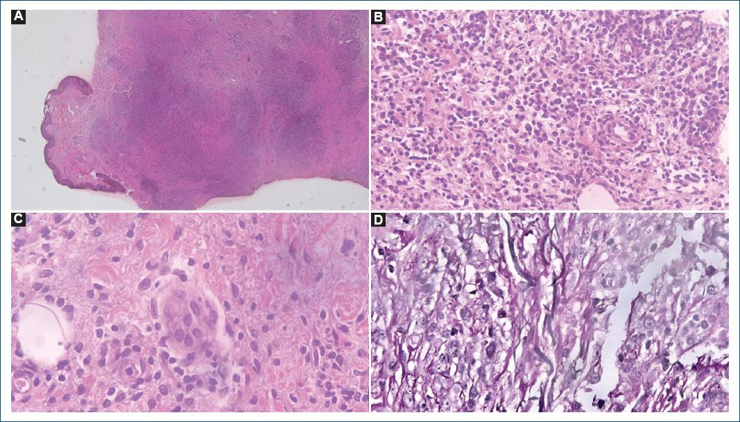
Figure 2 Hematoxylin-eosin-stained sections at ×40, ×200 and ×400, respectively. A: dense granulomatous infiltrate affecting the entire dermis and subcutaneous tissue. B: atypical lymphocytes, neutrophils, histiocytes, plasma cells, eosinophils composing the granuloma. C: scarce multinucleated giant cells. D: verhoeff-stained section at ×600; reduction of elastic fibers. 

Figure 3 Immunohistochemistry showing atypical cells expressing: A: CD3+; B: CD4+. 

GSS is a rare variant of mycosis fungoides that mainly affect caucasian men between the third and fifth decades of life1,2.

Histology shows, in addition to elastophagocytosis and emperipolesis, a granulomatous infiltrate mainly composed by atypical lymphocytes, macrophages, and multinucleated giant cells with 20-30 nuclei1-3. This last aspect was not found in our patient and to the best of our knowledge, only another similar case has been reported4. Regarding immunohistochemistry, it demonstrates a cell with a T-helper immunophenotype1, as in the present case, with monoclonal rearrangement of T-cell receptor genes in most tested patients1,2. In addition, some subpopulations of macrophages secrete metalloproteinases that are considered responsible for the degradation and remodeling of the dermal tissue3.

Furthermore, other lymphoproliferative disorders may be present in up to 50% of cases, so patients must be screened and followed up1,2. Due to its rarity, there is no standard treatment, and a complete remission of the disease has seldom been reported4-6.

References

1. Calonje E, Brenn T, Lazar AJ, Billings SD. Mckee's Pathology of the Skin with Clinical Correlations. 5th ed. Amsterdam:Elsevier;2019. [ Links ]

2. Roberti MR, Tuma CA. Granulomatous slack skin-a case report. An Bras Dermatol. 2007;82:445-9. [ Links ]

3. Feng Y, Wang S, Xie J, Ding B, Wang M, Zhang P, et al. Spatial transcriptomics reveals heterogeneity of macrophages in the tumor microenvironment of granulomatous slack skin. J Pathol. 2023;261:105-19. [ Links ]

4. Kempf W, Ostheeren-Michaelis S, Paulli M, Lucioni M, Wechsler J, Audring H, et al. Granulomatous mycosis fungoides and granulomatous slack skin:a multicenter study of the Cutaneous Lymphoma Histopathology Task force Group of the European Organization for Research and Treatment of Cancer (EORTC). Arch Dermatol. 2008;144:1609-17. [ Links ]

5. Martínez-Escala ME, González BR, Guitart J. Mycosis fungoides variants. Surg Pathol Clin. 2014;7:169-89. [ Links ]

6. Battesti G, Ram-Wolf C, Dobos G, Aubin F, Algros MP, Guenova E, et al. Granulomatous slack skin:clinical characteristics, prognosis and response to therapy. A study from the Cutaneous Lymphoma French Study Group. Br J Dermatol. 2022;187:790-3. [ Links ]

FundingNone.

Ethical disclosures

Protection of human and animal subjects. The authors declare that no experiments were performed on humans or animals for this study.

Confidentiality of data. The authors declare that they have followed the protocols of their work center on the publication of patient data.

Right to privacy and informed consent. The authors have obtained the written informed consent of the patients or subjects mentioned in the article. The corresponding author is in possession of this document.

Use of artificial intelligence for generating text. The authors declare that they have not used any type of generative artificial intelligence for the writing of this manuscript nor for the creation of images, graphics, tables, or their corresponding captions.

Received: May 30, 2023; Accepted: August 27, 2023

*Correspondence: Laísa E. de Hollanda E-mail: laisahollanda@gmail.com

Conflicts of interest

None.

Creative Commons License Portuguese Society of Dermatology and Venereology. Published by Permanyer. This is an open access article under the CC BY-NC-ND license