




 Inglês (pdf)
 Artigo em XML
 Referências do artigo

 Tradução automática
 Enviar este artigo por email
 Citado por SciELO
 Similares em
SciELO

 Permalink

Introduction
Historically, endoscope-associated infection was deemed to be very rare and the majority of documented cases were believed to be caused by noncompliance with guidelines [1, 2]. Recently, infection risk associated with contaminated, patient-ready flexible endoscopes has been reappraised [3] and several outbreaks of multidrug-resistant organisms have been particularly concerning [1].
A key recommendation is from scientific societies for reducing infection transmission risk focus in the culture of patient-ready endoscopes to detect contamination with organisms of concern [1, 4]. In fact, microbiological surveillance is a tool that we can use to monitor and adapt reprocessing protocols and a quality control measure for this process [2, 5-7], which allows us to identify endo-scopes with persistent contamination, despite their reprocessing [8].
Microbiological surveillance can also alert to possible defects in endoscopes and washer-disinfectors, so that endoscopy units can react at an early stage [9]. However, to implement an endoscope microbiological surveillance program, there is a need for specialized human and technical resources, both for sample collection and specimen culture and analysis [8]. Moreover, we must also consider the impact on procedure costs and environmental footprint. All these factors must be taken into account when developing microbiological surveillance protocols in endoscopy units, for them to be feasible in clinical practice.
Protocols for endoscope microbiological surveillance are established in several countries but lack standardization [4, 9]. There are significant variations in the timing and methods of sampling, the number of channels checked, the type of sampling solution used, the microbiological methods used (e.g., filtration vs. centrifugation), and the interpretation of the results [9]. The aim of this report was to summarize the critical aspects of the microbiological surveillance protocol drawn up by the Gastroenterology and the Clinical Pathology Departments in a Portuguese tertiary care academic Hospital.
Methods/Design Microbiological
Surveillance Protocol
The protocol resulted from a multidisciplinary partnership taking into account current scientific societies guidelines and the existing technical and human resources available in our hospital. Decisions were made regarding the frequency, sampling and culture methods and analysis. A microbiology request form was drawn up, accounting for the specificities of these samples (Table 1).
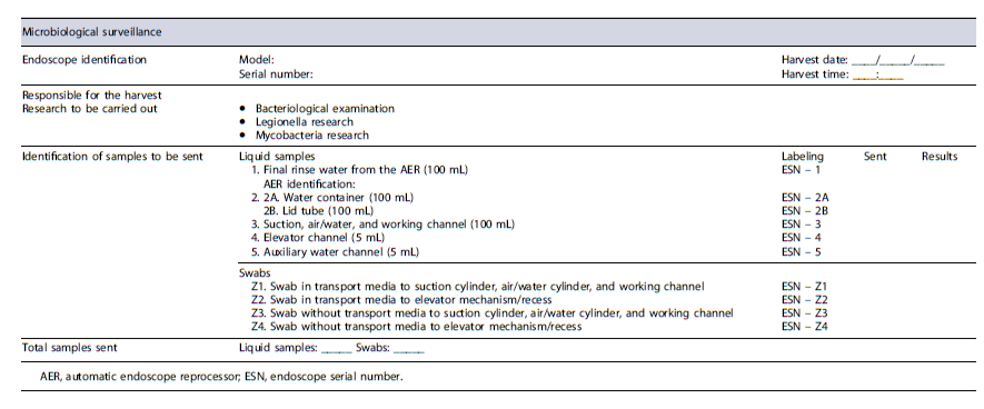
Sample Collection
The sample collection methods depend on endoscope type and model (Table 2). The collected samples are sterile saline liquid samples, as well as swabs (with and without transport media). No neutralizing substances were used, and the samples were transported to the local laboratory within a 30 min timeframe. Liquid samples are taken from the suction, air/water and biopsy channels of the endoscope, regardless the type and model. Then, depending on the presence of other channels, additional samples are obtained, such as from the elevator channel for duodenoscopes and curvilinear echoendoscopes, as well as from the auxiliary water channel in endoscopes that include this channel (colonoscope, gastroscope, pediatric colonoscope, or therapeutic endoscope) and balloon channel in the enteroscope (depending on the endoscope manufacturer). Additionally, samples of the final rinse water and from the water bottle are also collected.
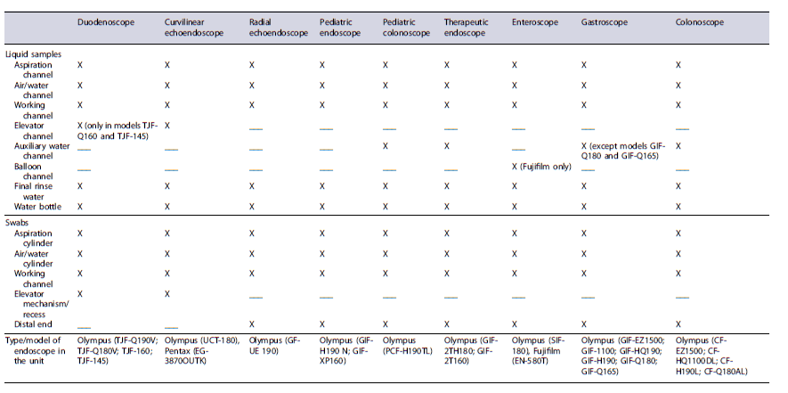
Regarding swabs, samples are collected from the suction and air/water cylinder and biopsy channel of all endoscopes. For endoscopes that include an elevator channel, an additional specimen is taken from the elevator mechanism and from the elevator recess. For other endoscope types, this is replaced by a sample from the external distal end of the endoscope. These samples are collected in duplicate swabs (with and without transport media).
For effective detection of microbial contamination in patient-ready endoscopes, an aseptic technique is used. First, a 100 mL sample of the final rinse water from the automatic endoscope reprocessor (AER) machine is taken. Then, from the water bottle used in the endoscopic procedure, two samples are taken: one directly from the water in the container (100 mL) and the other by irrigating sterile saline through the line of the tube lid (100 mL). These samples are placed in separate containers.
For endoscopes, swabs and liquid samples are collected. Swabs are soaked in sterile saline and only then, the sample is obtained from the suction aspiration cylinder, air/water cylinder, working channel, and elevator mechanism/recess in the case of duodenoscopes and linear echoendoscopes or from the distal end in the case of endoscopes that do not have an elevator mechanism (Fig. 1a). This is done for both swabs with and without transport media. The swab of the suction cylinder, air/water cylinder, and working channel is different from the swab of the elevator mechanism/recess or distal end of the endoscope, so that, in the event of a positive microbiological test, we can identify the microbiological contamination origin. For the liquid samples, 100 mL specimens of sterile saline from the suction channel, air/water channel, and working channel are taken using sterile connectors (Fig. 1b). Halfway through sample collection (after collecting 50 mL), we insert a previously sterilized brush through the suction channel and the working channel and cut the brush to be sent for analysis, together with the liquid sample, in order to improve sensitivity (“flush-brush-flush” method - Fig. 1c). The brush follows a sterilization process with ethylene oxide, as previously described by Ji et al. [10]. This sample is placed in a sterile container.
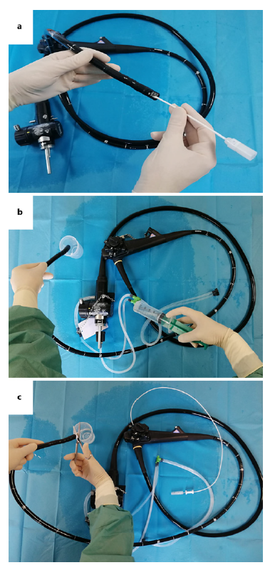
Fig. 1. Collection of microbiological samples. a Swab from the elevator recess from duodenoscope/echoendoscope (or from the distal end in the case of endoscopes that do not have an elevator). b Liquid sample from the aspiration, air/water, and working channel. c “Flush-brush-flush” method.
In the case of duodenoscopes or echoendoscopes with an elevator channel, or endoscopes with an auxiliary water channel, a 5 mL sample of sterile saline is irrigated through these channels, and it is sent for culture. These samples are placed in different containers.
The sample collection protocol for the duodenoscope is re-ported in the online supplementary material 1 (for all online suppl. material, see https://doi.org/10.1159/000539455). For other types of endoscopes, the protocol must be altered depending on the presence of other channels, such as the auxiliary water channel or balloon channel.
Microbiology Specimen Processing
The samples are processed immediately on arrival at the laboratory. In order to improve microbial recovery, samples with an optimum volume of 100 mL, as well as all samples over 5 mL, are previously concentrated by centrifugation for 15 min at 3,000 rpm. Subsequently, the samples are inoculated into a set of culture media suitable for the growth of the microorganisms to be assessed.
For the bacteriological test, 0.1 mL of each sample is inoculated using the 3-quadrant streak method onto Blood Agar (BioMérieux, Marcy-l’Étoile,France),andincubatedat 35°ina CO2 atmosphere for 5 days. For the detection of Legionella species, 0.1 mL of each sample is inoculated using the 3-quadrant streak method onto Buffered Charcoal Yeast Extract (BCYE; Oxoid, Madrid, Spain) and Glycine Vancomycin Polymyxin Cicloheximide (GVPC; Oxoid, Madrid, Spain) media and incubated at 35° in a CO2 atmosphere for 10 days. For the recovery of mycobacteria, 0.5 mL are inoculated into Mycobacteria Growth Indicator Tube (MGIT; Becton Dickinson, NJ, USA), with Middlebrook 7H9 broth, and incubated and monitored for 42 days using the BD BACTEC MGIT 960 system (Becton Dickinson, New Jersey, USA).
Bacteriological and Legionella testing of swab with transport medium samples is carried out by direct inoculation into the culture media and incubation conditions described above. To improve mycobacteriological recovery, the swabs without transport media for this test are previously embedded in MGIT and incubated at 35°C for 24 h, after which the MGIT is incubated under the conditions described for the liquid samples.
Frequency of Surveillance
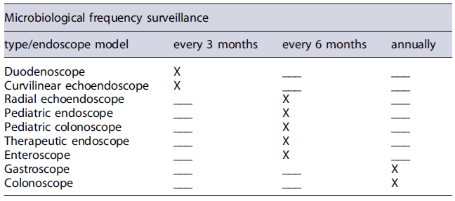
The timing and frequency for microbiological surveillance are determined by the type of endoscope (Table 3). Endoscopes that include an elevator channel are sampled every 3 months. For specific endoscopes, such as radial echoendoscopes, pediatric endoscopes/colonoscopes and therapeutic (dual channel) endoscopes, we collect samples every 6 months and, for gastroscopes and colonoscopes, annual surveillance is performed.
Timing of Sample Collection
Due to the personnel’s working hours, our protocol includes collecting samples within four to 6 h after reprocessing.
Results Report
As the microbiological control is performed under sterile conditions, any microbial growth is valued. In the presence of microbial growth, most of the species are identified using mass spectrometry on the VITEK® MS automated system (BioMérieux, Marcy-l’Étoile, France), based on MALDI-TOF (Matrix Assisted Laser Ionisation Time-of-Flight) technology. Mycobacteria are identified by molecular biology using GenoType Mycobacterium CM/AS (Hain Lifescience, Nehren, Germany), based on the PCR reaction and detection by DNA-strips reverse hybridization and an enzymatic staining reaction.
Discussion
The aim of microbiological surveillance was to ensure the quality of endoscope reprocessing, to identify deficiencies at an early stage and to provide information about possible risks [9]. The European Society of Gastrointestinal Endoscopy (ESGE) and the European Society of Gastroenterology and Endoscopy Nurses and Associates (ESGENA) provide guidance on how to perform endoscope microbiological surveillance, namely regarding frequency, sampling, sample volume, and culture, for either manual or automated reprocessing. If reprocessing is carried out automatically, the endoscope must be analyzed at the same time as the AER and the water bottle used in the procedure, in order to identify the source of the infection, in the event of a positive microbiological control [5, 11]. The ESGE/ESGENA guidelines recommend implementing microbiological surveillance on all endoscopes [5], while the US Food and Drug Administration (FDA), the Centers for Disease Control and Prevention (CDC), and the American Society for Microbiology (ASM) recommend it only for duodenoscopes [8].
The microbiological surveillance protocols used in different institutions differ in the methods of sampling, number of sampled channels, type and volume of sampling solutions and culture methods [1]. Considering the different guidelines and taking into account the resources available in our institution, we developed a protocol that could be implemented in our hospital, respecting its routines and taking into consideration sustainability issues.
In order to reduce the number of liquid samples from each endoscope, it was decided to combine certain samples in the same container, taking care not to interfere with the interpretation and analysis of the results or the corrective actions to be implemented, in the case of a positive control. Indeed, samples from different locations were combined ensuring that, in case of positive results, the following corrective measures were similar. As such, samples from the suction channel, air/water channel, and working channel were combined in the same container. Samples of additional channels, such as the elevator channel, auxiliary water channel, or balloon channel, are taken into another container, as well as samples from the final rinse water and water bottle in separate containers.
Regarding swabs, we collect samples from the suction cylinder, air/water cylinder, and working channel with the same swab and then use a second swab to sample the elevator mechanism/recess in duodenoscopes and linear echoendoscopes or in the case of other endoscopes, this is replaced by a sample from the distal end of the endoscope. It was decided to replace and not add this sample, in order not to increase the number of swabs needed from each endoscope and, once again, not to invalidate the implementation of the protocol in clinical practice. Therefore, our protocol includes collecting four swabs for each endoscope (two in transport media and two without transport media), in order to meet the laboratory capacities and reduce costs. At our endoscopy unit, we use sterile saline for irrigation through the water pump. Besides avoiding contamination, the use of sterile saline is intended to maintain an optimal environment for electric conductivity when performing polypectomy/mucosectomy. The saline vials are “single-day use”; therefore, they are discarded at the end of each shift. Thus, we opted to not include samples from these vials in our protocol. Nevertheless, for endoscopy units using reusable vials for water instillation through the water pump it would be relevant to include samples both from the vial as well as from the water itself.
For liquid sample collection, we selected the “flush-brush-flush” method. It is recommended by the CDC and was shown to be a more sensitive sampling technique in a Chinese study published in 2021 when compared with the conventional “flush method” (91.8 vs. 81.6% qualification rate) [8, 10]. No debris was found during sample collection.
With respect to sampling frequency, the recommendations are controversial, particularly concerning duodenoscopes. The ESGE/ESGENA guidelines recommend duodenoscope microbiological surveillance to be performed at least every 3 months, while, e.g., the World Gastroenterology Organization, in its recently published guideline, recommends it to be carried out monthly [5, 12]. A joint position statement by several Italian societies recommends monthly monitoring of duodenoscopes or after every sixty procedures [4].
Regarding frequency, and in order to be able to include all endoscopes in the surveillance program, we have chosen to follow the European recommendations, taking samples every 3 months from duodenoscopes and cur-vilinear echoendoscopes, and, at least, annually for other endoscope types, with specific endoscopes such as pediatric, therapeutic, enteroscopes, and radial echoendoscopes being sampled twice a year. With this frequency, we can conciliate the microbiology laboratory response with the inclusion of all endoscopes in use at our unit in the surveillance program, with the possibility for repeating sampling on endoscopes with positive results, after the implementation of corrective measures.
Our protocol has some limitations. First, our protocol focuses on the timing for sample collection. The Italian multisociety position paper recommends sampling to be performed at least 6 to 12 h after storage, to increase the likelihood of biofilm growth [4]. However, at our institution, we have only been able to collect samples within four to 6 h after reprocessing, due to the working hours of the departments and the professionals involved in sample collection, culture, and analysis. This fact can significantly impact on culture results, and in future studies we plan to evaluate this variable also taking into account endoscope storage conditions. With respect to the eluent solution used for sample collection, the fact that we do not use a buffered solution may be pointed as another limitation that can impact microorganism recovery and viability. In order to overcome this limitation, the samples are sent to the laboratory immediately after collection because it is known that delays in sample delivery and culturing can negatively interfere with the results.
In conclusion, endoscopy units should have detailed protocols for microbiological surveillance of their endoscopes as this is considered a structural quality indicator, to ensure high reprocessing quality [7]. These protocols should be drawn up by a multidisciplinary team that includes endoscopy nurses, gastroenterologists, microbiologists, and the antimicrobial stewardship team, guided by scientificsociety’s recommendations but adapted to each institution resources. The endoscopy units must establish a strong partnership with the microbiology laboratory and endoscope manufacturers, so that these protocols can be applied in clinical practice to guarantee patient safety.

