SciELO - Scientific Electronic Library Online

 
vol.82 número1Um caso de melanose de Becker na fronte: uma apresentação rara índice de autoresíndice de assuntosPesquisa de artigos
Home Pagelista alfabética de periódicos  

Serviços Personalizados

Journal

Artigo

Indicadores

Links relacionados

  • Não possue artigos similaresSimilares em SciELO

Compartilhar


Portuguese Journal of Dermatology and Venereology

versão impressa ISSN 2795-501Xversão On-line ISSN 2795-5001

Port J Dermatol Venereol. vol.82 no.1 Lisboa mar. 2024  Epub 06-Mar-2024

https://doi.org/10.24875/pjdv.23000071 

DERMATOLOGY IMAGES

Radiation-induced morphea: an uncommon entity

Morfeia pós-irradiação: uma entidade rara

Miguel Santos-Coelho1  *  https://orcid.org/0000-0002-2288-7306

Joana A. Barbosa1  https://orcid.org/0000-0001-7351-5302

Rodrigo Araújo-Carvalho1  https://orcid.org/0000-0002-3228-2482

1Department of Dermatology and Venereology, Hospital de Santo António dos Capuchos, Centro Hospitalar Universitário de Lisboa Central, E.P.E., Alameda Santo António dos Capuchos, Lisbon, Portugal


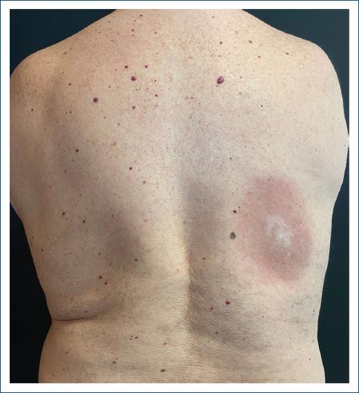
A 70-year-old man presented to the dermatology department with a slowly growing, asymptomatic, indurated 10 × 8 cm plaque, with a central white area and erythematous borders, of the right dorsal area (Fig. 1). Seventeen years earlier, the patient had developed a primary cutaneous follicle center lymphoma of the same region and had undergone treatment with 25 sessions of radiotherapy, with a complete response and no recurrence. Before this, no significant atrophy or radiodermatitis was noted.

Figure 1 Indurated plaque of the right dorsal area. 

Histopathology from a punch biopsy revealed no epidermal changes, a marked thickening of collagen fibers, with a dense, mainly perivascular and periadnexal, lymphohistiocytic infiltrate of the reticular dermis and hypodermis (Fig. 2). Perieccrine fat substitution by fibrosis was also present. These findings supported the diagnosis of radiation-induced morphea (RIM). The patient started topical betamethasone with gradual improvement over the next few months.

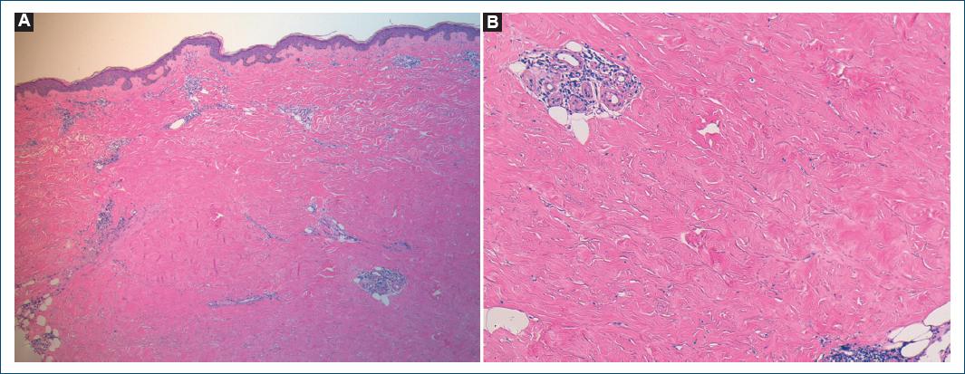
Figure 2 Punch biopsy revealing marked thickening of collagen fibers and a dense dermal perivascular and periadnexal lymphohistiocytic infiltrate (A: H and E, ×40; B: H and E, ×100). 

RIM is a rare entity most commonly associated with radiotherapy following breast cancer1. Most cases develop in the months following treatment, but a latent period of several years has been described2,3.

The precise pathophysiological mechanisms leading to RIM have not been established, but increased transforming growth factor-b signaling is thought to be a key element in inducing extracellular matrix deposition and extensive fibrosis2.

Differential diagnosis is vast and includes chronic radiation dermatitis, radiation recall dermatitis, and tumor recurrence, making skin biopsy an important step in these patients' evaluation.

Treatment is difficult and includes potent and superpotent topical corticosteroids, topical calcineurin inhibitors, oral methotrexate, oral corticosteroids, and phototherapy.

References

1. Machan A, Oumakhir S, Khalidi M, Hjira N, Boui M. Radiation-induced morphea:autoimmunity as a risk factor. Neth J Med. 2019;77:29-31. [ Links ]

2. Spalek M, Jonska-Gmyrek J, Gałecki J. Radiation-induced morphea-a literature review. J Eur Acad Dermatol Venereol. 2015;29:197-202. [ Links ]

3. Laetsch B, Hofer T, Lombriser N, Lautenschlager S. Irradiation-induced morphea:x-rays as triggers of autoimmunity. Dermatology. 2011;223:9-12. [ Links ]

FundingNone.

Ethical disclosures

Protection of human and animal subjects. The authors declare that no experiments were performed on humans or animals for this study.

Confidentiality of data. The authors declare that they have followed the protocols of their work center on the publication of patient data.

Right to privacy and informed consent. The authors have obtained the written informed consent of the patients or subjects mentioned in the article. The corresponding author is in possession of this document.

Use of artificial intelligence for generating text. The authors declare that they have not used any type of generative artificial intelligence for the writing of this manuscript nor for the creation of images, graphics, tables, or their corresponding captions.

Received: September 07, 2023; Accepted: September 17, 2023

*Correspondence: Miguel Santos-Coelho E-mail: mscoelho.derma@gmail.com

Conflicts of interest

None.

Creative Commons License Portuguese Society of Dermatology and Venereology. Published by Permanyer. This is an open access article under the CC BY-NC-ND license