SciELO - Scientific Electronic Library Online

 
vol.82 issue1Panniculitis in reactivation of Chagas disease in a cardiac transplant patientPrimary cutaneous anaplastic large-cell lymphoma: case report author indexsubject indexarticles search
Home Pagealphabetic serial listing  

Services on Demand

Journal

Article

Indicators

Related links

  • Have no similar articlesSimilars in SciELO

Share


Portuguese Journal of Dermatology and Venereology

Print version ISSN 2795-501XOn-line version ISSN 2795-5001

Port J Dermatol Venereol. vol.82 no.1 Lisboa Mar. 2024  Epub Jan 03, 2024

https://doi.org/10.24875/pjdv.23000073 

CASE REPORT

A case of autoimmune progesterone dermatitis

Um caso de dermatite autoimune à progesterona

Ana G. Lopes1  *  https://orcid.org/0000-0003-1103-1075

Catarina Cerqueira1 

Carlos Nogueira1 

Joana Gomes1 

Celeste Brito1 

1Department of Dermatology, Hospital de Braga, Braga, Portugal


Abstract

A 41-year-old female patient presents to the dermatology department with a 3-year history of a monthly relapsing pruritic eruption. These lesions appear 5-7 days before the onset of menses and resolve 3-4 days after menstruation. During her two previous pregnancies, she had no symptoms. She had been previously treated with antihistamines and oral corticosteroids with only temporary relief. On examination, during the luteal phase, the patient presented multiple maculopapular pruritic wheals distributed throughout the body. Several laboratory studies were performed and were all normal or negative, including auto-antibodies tests and hormonal analysis. Patch tests with the standard series of the Portuguese Contact Dermatitis Group, corticosteroid series, and metal series revealed positive reactions to nickel sulfate (++) and palladium chloride (+) at 72 h. An intradermal test with medroxyprogesterone at concentrations of 0.1 and 10 mg/mL was performed on the 7th day of the menstrual cycle. The test was positive 2 h after the injection and persisted for 24 h. The diagnosis of autoimmune progesterone dermatitis was made and the patient started tamoxifen 40 mg/day, with almost complete clinical clearing. Four months after, the dose was reduced, with no relapsing. Six months later, the patient remains free of symptoms.

Keywords Autoimmune progesterone dermatitis; Progesterone; Autoimmune urticaria; Urticaria; Intradermal test

Resumo

Descreve-se o caso de uma doente do sexo feminino de 41 anos, previamente saudável, sem antecedentes de dermatite atópica ou dermatite alérgica. A doente é avaliada na consulta de dermatologia por uma erupção pruriginosa recorrente, mensal, com 3 anos de evolução. Essas lesões geralmente apareciam 5 a 7 dias antes do início da menstruação e desapareciam aproximadamente 3 a 4 dias após o período menstrual. A doente referiu que quando esteve 2 vezes grávida e não teve sintomas nesse período. Ela tinha sido previamente tratada com anti-histamínicos e corticosteroides orais, com apenas alívio temporário. Ao exame físico, durante a fase lútea do período menstrual, a doente apresentava múltiplas maculopápulas eritematoedematosas, pruriginosas, distribuídas por todo o corpo. Vários estudos laboratoriais foram realizados e todos estavam normais ou negativos, incluindo testes de autoanticorpos e análises hormonais. Testes epicutâneos com a série padrão do Grupo Português de Dermatite de Contato (GPEDC), série de corticosteróides e série de metais revelaram reações positivas ao sulfato de níquel (++) e cloreto de paládio (+) às 72 horas. Foi realizado um teste intradérmico com medroxiprogesterona nas concentrações de 0.1 e 10 mg/mL no 7o dia do ciclo menstrual. O teste foi positivo 2 horas após a injeção e persistiu por 24 horas. Foi feito o diagnóstico de dermatite autoimune à progesterona e a doente foi tratada com tamoxifeno 40 mg/dia, com praticamente completa resolução clínica. Quatro meses após, a dose foi reduzida, sem recaída. Seis meses depois a doente continua sem sintomas ou efeitos adversos.

Palavras-chave Dermatite autoimune à; progesterona; Progesterona; Urticária autoimune; Urticária; Teste intradérmico

Introduction

Autoimmune progesterone dermatitis (APD) is a rare, cyclical, and mucocutaneous hypersensitivity reaction to peak levels of endogenous progesterone seen in women, in the luteal phase of the menstrual cycle. It is an underdiagnosed, complex disease associated with high morbidity1. Therefore, recognition of this process is important as it can result in significant quality of life impairment among women.

Our case report describes one of the rare cases of APD, manifesting as urticarial lesions.

Clinical case

We describe the case of a 41-year-old female patient, previously healthy, with no history of atopic or allergic dermatitis. She was not taking any medication and there was no history of atopic or allergic contact dermatitis.

The patient presented to the dermatology department with a 3-year history of a monthly relapsing pruritic eruption. The patient stated these lesions usually appeared 5-7 days before the onset of menses and resolved approximately 3-4 days after menstruation. She had been previously treated with antihistamines and oral corticosteroids with only temporary relief.

She had had no symptoms during her two pregnancies and she used an intrauterine copper device as a birth control method. There was no history of dysmenorrhea, menstrual irregularities, or oral contraceptive use.

On examination, during the luteal phase, the patient presented multiple maculopapular pruritic non-evanescent wheals distributed symmetrically throughout the body, lasting for more than 24 h, compatible with urticarial lesions.

Several laboratory studies were performed and were all normal or negative, including: ANA, anti-DNA, Sm, SSa, SSb, and RNP antibodies; C3, C4, CH100, and C1-inhibitor; thyroid stimulating hormone, T3, and T4; immunoglobulin G, immunoglobulin A, immunoglobulin M, and immunoglobulin E (IgE).

Hormonal analysis collected at day 3 and day 21 of the menstrual cycle revealed progesterone and estradiol serum levels were also normal during the follicular (1.57 ng/mL and 49.8 pg/mL, respectively) and the luteal phase (2.65 ng/mL and 64.3 pg/mL, respectively).

With the suspicion of APD an intradermal test with 0.1 mL of an 150 mg/mL aqueous solution of medroxyprogesterone serially diluted with 0.9% sodium chloride to concentrations of 0.1 and 10 mg/mL was performed on the 7th day of the cycle. A positive reaction was observed within 2 h, remaining positive for about 24 h (Figs. 1 and 2).

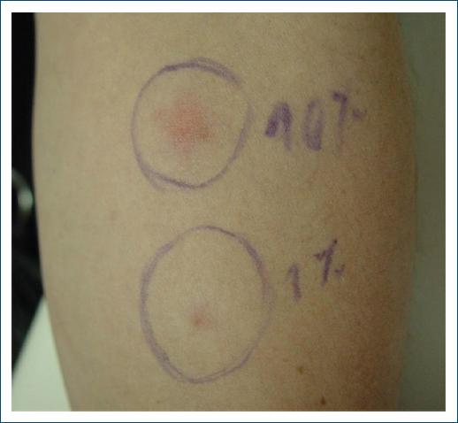
Figure 1 Intradermal test with aqueous solution of medroxyprogesterone (concentrations of 0.1 and 10 mg/mL) 2 h after. 

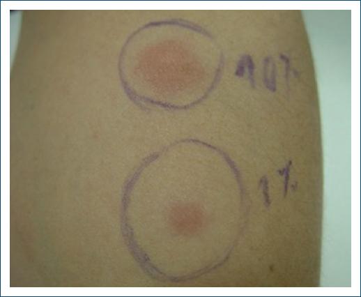
Figure 2 Intradermal test with aqueous solution of medroxyprogesterone (concentrations of 0.1 and 10 mg/mL) 5 h after. 

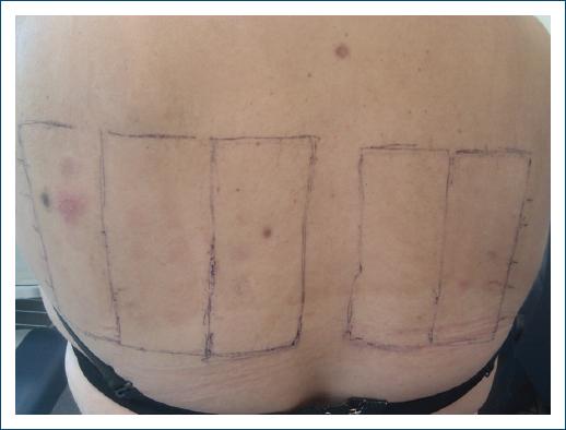
Patch tests were also performed, with the same medroxyprogesterone solution, the standard series of the Portuguese Contact Dermatitis Group, corticosteroids series, and metal series, which revealed positive reactions only to nickel sulfate 5% pet (++) and palladium chloride 1% pet (+) (Fig. 3).

Figure 3 Patch testing. 

The diagnosis of APD was made and the patient was treated with tamoxifen 40 mg/day, with almost complete clinical clearing. Four months after the dose was reduced to 20 mg/day, with no relapsing and subsequently to 10 mg/day. Six months later, the patient remains free of symptoms and with no side effects.

Discussion

APD is an extremely rare disease, characterized by recurring dermatologic manifestations during the luteal phase of the menstrual cycle2. APD has also been reported to be triggered by exogenous progesterone exposure or pregnancy, with peripartum onset and flares in a subset of patients3. In this case, the patient had no symptoms while she was pregnant.

The cause of APD is not known. It seems exogenous progesterone exposure, such as those used for oral contraception pills or in vitro fertilization, is an important cause of morbidity and may stimulate the body to form progesterone-specific IgE antibodies, that cross-link activate mast cells resulting in APD4. However, not all patients with this disease have a history of exposure to exogenous progesterone, as in the case we just described.

APD can have many different presentations including recurrent and cyclical urticaria with or without angioedema, anaphylaxis, pruritus, and dermatitis. Other presentations include vesiculobullous disorders, erythema multiforme, fixed drug eruptions, aphthous stomatitis, maculopapular rash, and recalcitrant dermatitis5-7. The lesions are characteristically symmetrical and occur on the face, trunk, and extremities. Our patient presented with urticarial lesions, which is one of the most common manifestations of this dermatitis.

The mean age at the beginning of symptoms is 27.3 years6. Symptoms usually appear 3-4 days before menstruation when progesterone levels peak and resolve within a few days after the onset of menstruation as progesterone levels reduce, only to recur just before the next period1, as in this case.

Diagnosis is difficult and often delayed. It is frequently made based on the exclusion of all possible differential diagnoses.

The diagnostic criteria for APD proposed by Warin are: skin lesions related to menstrual cycle; symptomatic improvement after inhibiting progesterone secretion by suppressing ovulation; positive response to intradermal testing with progesterone8. Intradermal progesterone tests may be used to help diagnose APD; however, the test is not standardized, has unknown sensitivity and specificity, and test results do not typically change management. In a series of 24 cases of APD, only 50% of patients showed a positive result to this test. In patients presenting with urticaria and/or anaphylaxis, the intradermal skin test may potentially be of more value9. In this case, we performed an intradermal test with an aqueous solution of medroxyprogesterone at concentrations of 0.1 and 10 mg/mL on the 7th day of the menstrual cycle, and it was positive.

In regards to treatment, primary treatment includes prescribing a combination of oral contraceptives. Other successful treatment options include gonadotropin-releasing hormone agonists, danazol and tamoxifen; topical and oral antihistamines and steroids to treat cutaneous symptoms; and bilateral oophorectomy in patients experiencing persistent symptoms2. Here, the patient was treated with tamoxifen with complete resolution of symptoms.

Conclusion

The diagnosis of APD still remains a challenge, contributing to a significant delay in diagnosis, requiring further clarification of criteria and development of accurate diagnostic tests. Recognition of this rare condition needs a high index of suspicion.

References

1. Gonçalves DG, Kalil J, Giavina-Bianchi P. Dermatite autoimune àprogesterona. Arq Asma Alerg Imunol. 2017;1:357-62. [ Links ]

2. Irshad S, Haider MS, Master MF, Asif N, Khalil A. Autoimmune progesterone dermatitis. Cureus. 2021;13:e19217. [ Links ]

3. Aghazadeh N, Berry NA, Torgerson RR, Park MA, Davis DM. Autoimmune progesterone dermatitis:a retrospective case series. Int J Womens Dermatol. 2022;8:e009. [ Links ]

4. Buchheit KM, Bernstein JA. Progesterone hypersensitivity:heterogeneous manifestations with a common trigger. J Allergy Clin Immunol Pract. 2017;5:566-74. [ Links ]

5. Aghazadeh N, Chattha AJ, Hartz MF, Davis DM. Autoimmune progesterone dermatitis in the adolescent population. Pediatr Dermatol. 2021;38:380-4. [ Links ]

6. Nguyen T, Razzaque Ahmed A. Autoimmune progesterone dermatitis:update and insights. Autoimmun Rev. 2016;15:191-7. [ Links ]

7. Lee MK, Lee WY, Yong SJ. A case of autoimmune progesterone dermatitis misdiagnosed as allergic contact dermatitis. Allergy Asthma Immunol Res. 2011;3:141-4. [ Links ]

8. Warin AP. Case 2. Diagnosis:erythema multiforme as a presentation of autoimmune progesterone dermatitis. Clin Exp Dermatol. 2001;26:107-8. [ Links ]

9. Foer D, Buchheit KM, Gargiulo AR, Lynch DM, Castells M, Wickner PG. Progestogen hypersensitivity in 24 cases:diagnosis, management, and proposed renaming and classification. J Allergy Clin Immunol Pract. 2016;4:723-9. [ Links ]

FundingNone.

Ethical disclosures

Protection of human and animal subjects. The authors declare that no experiments were performed on humans or animals for this study.

Confidentiality of data. The authors declare that they have followed the protocols of their work center on the publication of patient data.

Right to privacy and informed consent. The authors have obtained the written informed consent of the patients or subjects mentioned in the article. The corresponding author is in possession of this document.

Use of artificial intelligence for generating text. The authors declare that they have not used any type of generative artificial intelligence for the writing of this manuscript nor for the creation of images, graphics, tables, or their corresponding captions.

Received: September 13, 2023; Accepted: October 31, 2023

*Correspondence: Ana G. Lopes E-mail: ana.gabriela.castro@hb.min-saude.pt

Conflicts of interest

None.

Creative Commons License Portuguese Society of Dermatology and Venereology. Published by Permanyer. This is an open access article under the CC BY-NC-ND license