SciELO - Scientific Electronic Library Online

 
vol.81 número4Uma variante histológica rara de sarcoma de KaposiAlopecia tracional índice de autoresíndice de assuntosPesquisa de artigos
Home Pagelista alfabética de periódicos  

Serviços Personalizados

Journal

Artigo

Indicadores

Links relacionados

  • Não possue artigos similaresSimilares em SciELO

Compartilhar


Portuguese Journal of Dermatology and Venereology

versão impressa ISSN 2795-501Xversão On-line ISSN 2795-5001

Port J Dermatol Venereol. vol.81 no.4 Lisboa dez. 2023  Epub 20-Dez-2023

https://doi.org/10.24875/pjdv.23000030 

DERMATOLOGY IMAGES

Atypical porphyria cutanea tarda mimicking morphea

Porfiria cutânea tarda atípica mimetizando morfeia

Maria J. Guimarães1  *  https://orcid.org/0000-0002-3247-2470

Carlos M. Nogueira1 

Catarina Cerqueira1 

Ana G. Lopes1 

Celeste Brito1 

Catarina Araújo1 

1Department of Dermatology and Venereology, Hospital de Braga, Braga, Portugal


A 65-year-old man with a history of alcohol abuse presented with cicatricial alopecia and whitish sclerotic plaques in the upper chest, distributed mainly on the V area of the lower neck and lower sternum (Fig. 1). In addition, physical examination revealed a hyperpigmented area of hypertrichosis on the interparietal and left parietal region (Fig. 2).

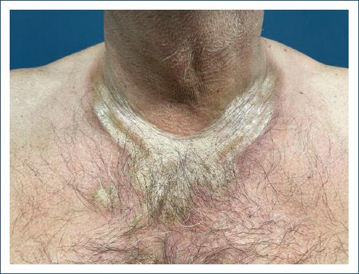
Figure 1 White-yellow atrophic plaques in the “V” area of the upper chest with a hyperpigmented border. 

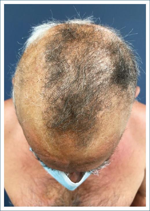
Figure 2 Alopecic patches on the scalp and hyperpigmented area of hypertrichosis on the interparietal and left parietal region. 

Laboratory evaluation showed iron overload and elevated transaminases. Antinuclear antibodies and infectious serologies, including borrelia, were negative. A cutaneous biopsy of the scalp and neck was compatible with morphea, and thus, narrow band ultraviolet B phototherapy was initiated.

One month later, the patient reported blistering and crusting of the forearms. Urine analysis revealed increased uroporphyrins establishing the diagnosis of porphyria cutanea tarda (PCT). The patient was started on bimonthly phlebotomies, and photoprotection and alcohol withdrawal were recommended.

Sclerodermiform changes have been reported in 2% of PCT patients1,2. Clinically, lesions may resemble morphea, presenting with hyperpigmentation rather than a peripheral lilac ring3,4. Scalp lesions may present as scarring alopecia, also called alopecia porphyrinica3. Isolated sclerodermiform changes, without the typical clinical picture of PCT, pose a diagnostic challenge. Histopathology cannot reliably distinguish morphea from sclerodermiform PCT4. Given the clinical and pathological resemblance, one must consider this alternative diagnosis, especially when facing therapy failure.

REFERENCES

1. Simon N, Korom I, Szekeres L, Morvay M, KószóF. Sclerodermiform porphyria. Z Hautkr. 1986;61:1607-21. [ Links ]

2. Friedman SJ, Doyle JA. Sclerodermoid changes of porphyria cutanea tarda:possible relationship to urinary uroporphyrin levels. J Am Acad Dermatol. 1985;13:70-4. [ Links ]

3. Volksbeck SI, Nashan D, Bruckner-Tuderman L, Braun-Falco M. Localized sclerosis of the scalp (alopecia porphyrinica) as predominant presentation of porphyria cutanea tarda. J Eur Acad Dermatol Venereol. 2007;21:1125-7. [ Links ]

4. Stevens HP, Ostlere LS, Rustin MH, Black CM. Generalized morphoea secondary to porphyria cutanea tarda. Br J Dermatol. 1993;129:455-7. [ Links ]

FundingNone.

Ethical disclosures

Protection of human and animal subjects. The authors declare that no experiments were performed on humans or animals for this study.

Confidentiality of data. The authors declare that they have followed the protocols of their work center on the publication of patient data.

Right to privacy and informed consent. The authors have obtained the written informed consent of the patients or subjects mentioned in the article. The corresponding author is in possession of this document.

Use of artificial intelligence for generating text. The authors declare that they have not used any type of generative artificial intelligence for the writing of this manuscript, nor for the creation of images, graphics, tables, or their corresponding captions.

Received: April 20, 2023; Accepted: July 26, 2023

*Correspondence: Maria J. Guimarães E-mail: mjcunhaguimaraes@gmail.com

Conflicts of interest

None.

Creative Commons License Portuguese Society of Dermatology and Venereology. Published by Permanyer. This is an open access article under the CC BY-NC-ND license