


 Inglês (pdf)
 Artigo em XML
 Referências do artigo
 Enviar este artigo por email
 Citado por SciELO
  Similares em
SciELO
Similares em
SciELO 
 Permalink
Our case focuses on a 30-year-old female patient with no relevant priors.
The patient was referred to our dermatology department due to the appearance of a cutaneous lesion on the left leg during her last pregnancy which was 6 months ago.
At the dermatologic examination, she presented a macule with brown pigment on the center and an erythematous halo, well-demarcated, with superficial scaling, < 1 cm in diameter, and on the anterior surface of the left leg (Figure 1).
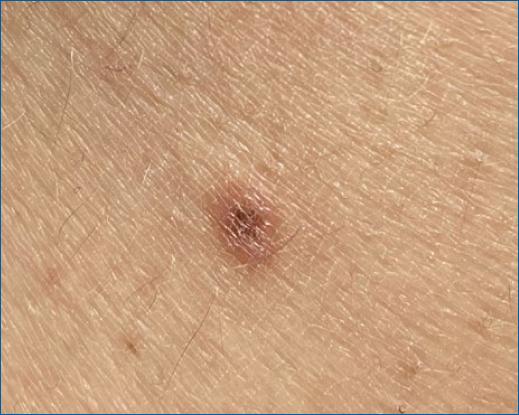
Figure 1 Macule with brown pigment on the center and an erythematous halo, well-demarcated, with superficial scaling, < 1 cm in diameter, and on the anterior surface of the left leg.
Dermoscopy revealed light brown dots and globules on a yellow background, with dotted vessels and white streaks at the periphery (Figure 2).
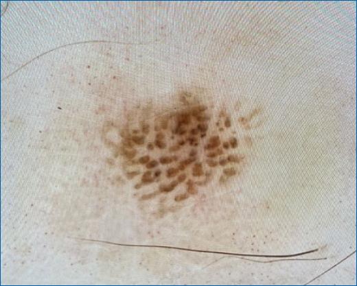
Figure 2 Dermoscopy of the lesion, showing light brown dots and globules on a yellow background, with dotted vessels, and white streaks at the periphery.
A cutaneous biopsy showed acanthosis with mild orthokeratotic hyperkeratosis, larger than usual keratinocytes, and hyperpigmentation of the basal layer. The histopathological findings were compatible with a large cell acanthoma (LCA).
An LCA is a rare epidermal benign tumor, considered by some a variant of the solar lentigo with cellular hypertrophy. It occurs most frequently in women, older people, and in sun-exposed sites, such as the face and extremities1,2. Clinically, LCA may be difficult to be differentiated from a solar lentigo, a pigmented actinic keratosis, or a flat seborrheic keratosis3. A recently published study performed on 33 lesions (26 patients) identified distinct dermoscopic findings of LCA4. The most frequent dermoscopic findings are a yellow opaque homogenous area, grey/brown dots and globules, a moth-eaten border, white streaks, and a pseudonetwork2,4, most of which were also present in this case. Another study evaluated 13 patients and also identified these as the most frequent dermoscopic features and found that milia-like cysts and white to yellow surface scale were uncommon findings5.
Therefore, dermoscopy is a noninvasive tool that can significantly aid in the diagnosis of LCA.

