SciELO - Scientific Electronic Library Online

 
vol.80 issue2Cutaneous phaeohyphomycosis caused by Exophiala dermatitidis in an immunosuppressed patient: A case report and brief reviewAsymptomatic erythematous depression on the thumb author indexsubject indexarticles search
Home Pagealphabetic serial listing  

Services on Demand

Journal

Article

Indicators

Related links

  • Have no similar articlesSimilars in SciELO

Share


Portuguese Journal of Dermatology and Venereology

Print version ISSN 2795-501XOn-line version ISSN 2795-5001

Port J Dermatol Venereol. vol.80 no.2 Lisboa June 2022  Epub Aug 02, 2022

https://doi.org/10.24875/pjd.m22000023 

CASE REPORT

Managing squamous cell carcinoma in recessive dystrophic epidermolysis bullosa with electrochemotherapy

Eletroquimioterapia em carcinoma espinocelular na epidermólise bolhosa distrófica recessiva

Mafalda Pestana1  *  https://orcid.org/0000-0003-1361-4995

Margarida Brito-Caldeira1 

Victor Farricha2 

Sara Carvalhal2 

Joana Bartolo2 

Cecília Moura2 

Joaninha Costa-Rosa2 

Emanuel Gouveia2 

Hugo Nunes2 

Mariluz Martins2 

1Dermatology and Venereology Department, Centro Hospitalar Universitário de Lisboa Central

2Skin Cancer Multidisciplinary Group, Instituto Português de Oncologia de Lisboa. Lisboa, Portugal


Abstract

Epidermolysis bullosa (EB) represents a group of congenital disorders caused by mutations in skin structural proteins. Squamous cell carcinoma (SCC) is one major complication and the leading cause of death of several subtypes of EB. We report a case of a 38-year-old female patient with recessive dystrophic EB that underwent electrochemotherapy (ECT) for treatment of cutaneous SCC in the right foot. Two sessions were performed, in which the intravenous administration of bleomycin was followed by the local application of electric pulses. We verified a partial response in 8th week of follow up and a complete response in the 24th week.

ECT is a local treatment for cutaneous and subcutaneous tumors, mainly as palliative care for metastases. Studies have shown its effectiveness in primary tumors that are unresectable due to size and location, and it has been proposed as a new treatment modality for SCCs in EB with high overall response.

Keywords Recessive dystrophic epidermolysis bullosa; Squamous cell carcinoma; Electrochemotherapy

Resumo

A epidermólise bolhosa (EB) representa um grupo de doenças congénitas causadas por mutações em genes que codificam proteínas estruturais da pele. O carcinoma espinocelular (CEC) é uma das suas complicações mais temidas, sendo a principal causa de morte nalguns subtipos de EB. Apresenta-se o caso de uma doente de 38 anos com EB distrófica recessiva, que foi submetida a eletroquimioterapia (EQT) para o tratamento de um CEC no pé direito. Foram realizadas duas sessões de tratamento, com administração endovenosa de bleomicina seguida da aplicação de pulsos elétricos. Verificou-se uma resposta parcial às 8 semanas de follow-up e resposta completa às 24 semanas.

A EQT é um tratamento local para tumores cutâneos e subcutâneos, primeiramente como modalidade paliativa em metástases. Estudos demonstram a sua eficácia no tratamento de tumores cutâneos primários irressecáveis, pelas suas dimensões ou localização, tendo sido proposta como modalidade terapêutica nos CECs na EB, com boa resposta global.

Palavras-chave Epidermólise bolhosa distrófica recessiva; Carcinoma espinocelular; Eletroquimioterapia

Introduction

Epidermolysis Bullosa (EB) represents a group of rare congenital disorders caused by genetic variants in skin structural proteins, resulting in disruption of the dermal-epidermal junction. It is a clinically and genetically heterogeneous disorder characterized by skin fragility, with a propensity to impaired wound healing and blister formation. Based upon the ultrastructural levels of skin cleavage, EB is classified into four major types: simplex, junctional, dystrophic, and Kindler Syndrome. Recessive dystrophic EB (RDEB) results from a mutation in the COL7A1 gene that encodes the alpha-1 chain of type VII collagen1,2.

Squamous cell carcinoma (SCC) is one of the most feared complications and the leading cause of death of several subtypes of EB. The risk is higher for RDEB, especially in the generalized severe form3. To date, surgery is the first-line treatment for EB-associated SCC (EB-SCC), however, these tumors show an aggressive course, with high rates of recurrence even following complete excision4,5. Systemic chemotherapy and radiotherapy are only recommended as palliative modalities since their side effects may overweight the benefits in these fragile patients6,7. Despite all treatments available, there is a high morbidity and mortality associated to EB-SCCs, with significantly reduced life expectancy.

Electrochemotherapy (ECT) is a local treatment for cutaneous and subcutaneous tumors. The first ECT clinical trial was performed in 1991 in Villejuif, France, on head and neck cutaneous metastatic SCCs8. The European Standard Operating Procedures of ECT (ESOPE) project, which established the guidelines for a safe application of ECT in clinical practice was published in 20069. Even though ECT is mainly applied as palliative care for metastases, it may also be used for primary tumors that are unresectable due to size and location10,11. ECT has been proposed as a new treatment modality for EB-SCCs, due to its effectiveness and favorable safety profile4,12,13.

Case Report

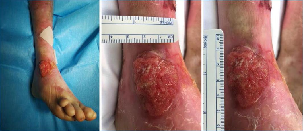
We report the case of a 38-year-old female patient with RDEB who presented with a cutaneous SCC in the dorsum of the right foot (Fig. 1). The lesion had been growing for the past 4 months, and was successfully treated with ECT, obviating the need for further treatments. The inclusion criteria and technical procedures followed the ESOPE9. Patient underwent an ECT session using the ePORE Gx device (Pes Med, Italy) in which bleomycin was administrated intravenously at a dose of 15 000 UI/m2 for 2 minutes, followed by electric pulses 8 minutes later. The lesion was examined and measured at follow-up appointments to evaluate the response to treatment and efficacy was assessed according to the Response Evaluation Criteria in Solid Tumours14. We reported stable disease 8 weeks after the first treatment, so we proposed a second session, which the patient consented. The second ECT session was performed 9 weeks after the first one, using the Cliniporator 1 device (IGEA, Italy). At 8 weeks of follow-up, we reported a partial response (Fig. 2a), with a complete response and skin healing of the treated area at 24 weeks of follow-up (Fig. 2b). ECT was well tolerated, with only mild adverse effects reported, which included muscle contractions during each pulse and local pain, erythema, and ulceration in the follow-up period. After the first three postoperative months, in which the patient was evaluated every two weeks, a quarterly follow-up was performed in the first year and every four months thereafter. At 64 weeks of follow-up, the patient is still free of recurrence.

Figure 1 Clinical presentation. An erosive tumor, 3 × 4 cm in size, localized in the dorsum of the right foot. 

Figure 2 Clinical response to treatment. A: a partial response was reported at 8 weeks of follow-up, with superficial ulceration at the site of the SCC. B: at 24 weeks of follow-up, a complete response was reported, with no clinically evident disease. 

Discussion

A major complication of EB is the development of multiple cutaneous SCCs, which generally develop in early adulthood and tend to arise at sites of chronic non-healing wounds or hyperkeratotic lesions. These tumors may be difficult to identify clinically since they frequently resemble areas of nonmalignant EB ulceration and wounds, so it is essential to maintain a straight clinical follow-up and have a low suspicion threshold to perform biopsy3,15. They represent the leading cause of death in EB at or after mid-adolescence, with death from cutaneous SCC occurring within 5 years of the diagnosis of the first SCC in most patients. The cumulative risk of at least one SCC in patients with generalized severe form of RDEB is 7.5%, 68%, and 90% by ages 30, 35, and 55 respectively. In contrast, the risk of these tumors is <25% by age 45 years in other forms of RDEB16.

The first line treatment for cutaneous EB-SCCs is surgery however these lesions tend to recur locally as their borders are usually indistinct4. Radiotherapy and conventional chemotherapy have been reserved as palliative modalities for advanced or locally recurrent SCC so other treatment options should be considered6,7. ECT combines the administration of a low dose of a chemotherapeutic agent, such as cisplatin or bleomycin, and the local application of short intense electric pulses. The electric fields transiently permeabilize the cell membrane, allowing the entrance of the drug into the neoplastic cells, enhancing its effectiveness and reducing its side effects. Only transient and mild adverse effects are usually reported8,10, as in the present case. Although it has no impact in the systemic progression of the disease, it has been proposed as a treatment modality for EB-SCC with high overall response.

In this case, we verified a complete response of a cutaneous SCC after two well tolerated sessions of ECT. This report aims to reinforce the potential of ECT as a viable approach for SCCs in patient with EB, without the high risk of functional impairment after surgical excision and no contraindications related to the disease.

What does this study add

We aim to reinforce the potential of electrochemotherapy as a viable approach for squamous cell carcinoma in patients with epidermolysis bullosa, without the high risk of functional impairment after surgical excision.

Prizes and previous presentations

This case has been presented as a clinical case on “Reunião de Primavera da Sociedade Portuguesa de Dermatologia e Venereologia”, Aveiro, 9–10th July 2021 and as an e-poster on “EADV 30th congress,” Virtual, 29th September–2nd October 2021.

REFERENCES

1. Gedde-Dahl T Jr. Epidermolysis bullosa. A clinical, genetic and epidemiologic study. Baltimore:Johns Hopkins University Press;1971. p. 1–180. doi:10.7326/0003-4819-75-4-659_2. [ Links ]

2. Intong LR, Murrell DF. Inherited epidermolysis bullosa:new diagnostic criteria and classification. Clin Dermatol. 2012;30(1):70–7. doi:10.1016/j.clindermatol.2011.03.012. [ Links ]

3. Condorelli A, Dellambra E, Logli E, Zambruno G, Castiglia D. Epidermolysis bullosa-associated squamous cell carcinoma:from pathogenesis to therapeutic perspectives. Int J Mol Sci. 2019;20(22):5707. doi:10.3390/ijms20225707. [ Links ]

4. Mellerio J, Robertson S, Bernardis C, Diem A, Fine JD, George R, et al. Management of cutaneous squamous cell carcinoma in patients with epidermolysis bullosa:best clinical practice guidelines. Br J Dermatol. 2015;174(1):56–67. doi:10.1111/bjd.14104. [ Links ]

5. Yamada M, Hatta N, Sogo K, Komura K, Hamaguchi Y, Takehara K. Management of squamous cell carcinoma in a patient with recessive type epidermolysis bullosa dystrophica. Dermatol Surg. 2004;30:424–1429. doi:10.1111/j.1524-4725.2004.30440.x. [ Links ]

6. Bastin KT, Steeves RA, Richards MJ. Radiation therapy for squamous cell carcinoma in dystrophic epidermolysis bullosa:case reports and literature review Am J Clin Oncol. 1997;20(1):55–8. doi:10.1097/00000421-199702000-00012. [ Links ]

7. Lentz SR, Raish RJ, Orlowski EP, Marion JM. Squamous cell carcinoma in epidermolysis bullosa:treatment with systemic chemotherapy. Cancer. 1990;66:1276–8. doi:10.1002/1097-0142(19900915)66:6<1276::aid-cncr2820660629>3.0.co;2-a. [ Links ]

8. Mir LM, Belehradek M, Domenge C, Orlowski S, Poddevin B, Belehradek J Jr, et al. Electrochemotherapy, a new antitumour treatment:first clinical trial. C R Acad Sci III. 1991;313(13):613–8. doi:10.1002/1097-0142(19931215)72:12<3694::aid-cncr2820721222>3.0.co;2-2. [ Links ]

9. Mir LM, Gehl J, Sersa G, Collins CG, Garbay JR, Billard V, et al. Standard operating procedures of the electrochemotherapy:instructions for the use of bleomycin or cisplatin administered either systemically or locally and electric pulses delivered by the Cliniporator by means of invasive or non-invasive electrodes. Eur J Cancer. 2006;4:14–25. doi:10.1016/j.ejcsup.2006.08.003. [ Links ]

10. Mali B, Jarm T, Snoj M, Sersa G, Miklavcic D. Antitumor effectiveness of electrochemotherapy:a systematic review and meta-analysis. Eur J Surg Oncol. 2013;39:4–16. doi:10.1016/j.ejso.2012.08.016. [ Links ]

11. Mir L, Glass L, Serša G, TeissiéJ, Domenge C, MiklavčičD, et al. Effective treatment of cutaneous and subcutaneous malignant tumours by electrochemotherapy. Br J Cancer. 1998;77(12):2336–42. doi:10.1038/bjc.1998.388. [ Links ]

12. Diociaiuti A, Rotunno R, el Hachem M, Latorre S, Cozza R, Curatolo P, et al. Electrochemotherapy, a potential new treatment for the management of squamous cell carcinoma in patients with recessive dystrophic epidermolysis bullosa:report of three cases. J Eur Acad Dermatol Venereol. 2015 Jul;30(7):1195–6. doi:10.1111/jdv.13116. [ Links ]

13. Bartolo J, Farricha V, Carvalhal S, Moura C, Abecasis N. Electrochemotherapy, a local treatment for squamous cell carcinoma in patients with recessive dystrophic epidermolysis bullosa. Dermatol Ther. 2020;33(6). [ Links ]

14. Therasse P, Arbuck SG, Eisenhauer EA, Wanders J, Kaplan RS, Rubinstein L, et al. New guidelines to evaluate the response to treatment in solid tumours. European Organization for Research and Treatment of Cancer, National Cancer Institute of the United States, National Cancer Institute of Canada. J Natl Cancer Inst. 2000;92:205–16. doi:10.1111/dth.14093. [ Links ]

15. Fine JD, Mellerio J. Extracutaneous manifestations and complications of inherited epidermolysis bullosa. Parts I&II. J Am Acad Dermatol. 2009;61:367–402. doi:10.1016/j.jaad.2009.03.053. [ Links ]

16. Fine JD, Johnson LB, Weiner M, Li KP, Suchindran C. Epidermolysis bullosa and the risk of life-threatening cancers:national EB registry experience, 1986–2006. J Am Acad Dermatol. 2009;60:203–11. doi:10.1016/j.jaad.2008.09.035. [ Links ]

FundingThe authors received no financial support for the authorship or publication of this article.

Ethical disclosures

Protection of human and animal subjects. The authors declare that no experiments were performed on humans or animals for this investigation.

Confidentiality of data. The authors declare that they have followed the protocols of their work center on the publication of patient data and that all the patients included in the study have received sufficient information and have given their informed consent in writing to participate in that study.

Right to privacy and informed consent. The authors have obtained the informed consent of the patients and/or subjects mentioned in the article. The author for correspondence is in possession of this document.

Received: October 25, 2021; Accepted: February 10, 2022

*Corresponding author: Mafalda Pestana E-mail: mafaldambp@gmail.com

Conflicts of interest

None to declare.

Creative Commons License Portuguese Society of Dermatology and Venereology. Published by Permanyer. This is an open access article under the CC BY-NC-ND license