


 English (pdf)
 Article in xml format
 Article references
 Send this article by e-mail
 Cited by SciELO
  Similars in
SciELO
Similars in
SciELO 
 Permalink
Case report
We report the case of a six-month-old male that presented with a complex dermatosis with evolution since birth. He was otherwise healthy, with normal mental developmental milestones for his age. The cutaneous findings were asymptomatic, and family history was unremarkable.
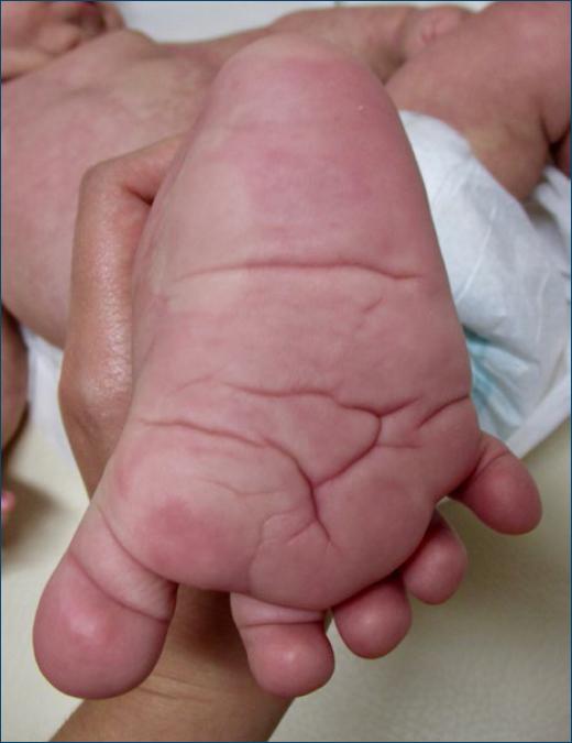
Physical examination revealed an extensive reticulated erythematous patch that occupied the plants bilaterally and extended superiorly to both legs (Fig. 1). The lesion also affected the trunk, namely the right anterior chest wall, where it showed sharp demarcation at the midline (Fig. 2). It further extended to the abdominal and dorsolumbar regions. In addition, the patient had macrocephaly and gigantism of the hands and feet, with an acral furrowing of the skin (Fig. 3). He also presented with a striking “sandal gap-toe” (an increased gap between the first and second toes). Although the right lower limb had a larger circumferential diameter compared to the left, both limbs had the same length. Later on, during follow-up, he developed vaguely papillomatous plaque lesions located at the right cervical region that extended to the right arm, suggestive of epidermal nevi.
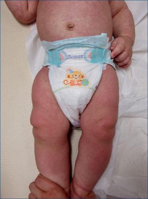
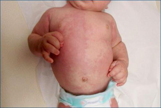
Figure 2 Reticulated erythematous patch of the right anterior chest wall, with a sharp demarcation at the midline.
Taking clinical picture into account, differential diagnosis of overgrowth syndrome was suggested. Lesional biopsy in the plantar region was performed, revealing multiple adipose tissue lobes accompanied by thick-walled vessels, numerous hypertrophied nerve bundles, and thickened collagen septa. These findings, although not specific, raise the hypothesis of hamartomatous hyperplasia. In addition, genetic testing was performed in lesional skin, revealing a PIK3CA mutation, and thus establishing the diagnosis of CLOVES syndrome. The patient remains under close monitoring by a multidisciplinary team of dermatology, neurology and orthopedics.
Discussion
PIK3CA-related overgrowth spectrum (PROS) designates a heterogeneous group of asymmetric overgrowth disorders caused by an activating somatic mutation in the PIK3CA gene1. These mutations consequently lead to an abnormal activation of the phosphatidylinositol-3-kinase-AKT-mammalian target of rapamycin (PI3K-AKT-mTOR) pathway. CLOVES syndrome is an exceedingly uncommon entity considered to be part of the PROS. Its acronym stands for congenital lipomatous overgrowth (CLO), vascular anomalies (V), epidermal nevi (E), and skeletal deformities (S). There is considerable overlap with other entities associated with PIK3CA mutations, namely Klippel–Trenaunay syndrome.
Lipomatous overgrowth is a key sign, usually involving the back and trunk1. It is present at birth, but their small size means that they occasionally go undetected until adulthood2. These lipomatous masses grow progressively over time, sometimes infiltrating nearby structures, namely the abdominal cavity, retroperitoneum, mediastinum, pleura and medullary cavity, with subsequent morbidity3.
Vascular anomalies are another prominent finding in CLOVES syndrome, with a wide range of clinical manifestations. More often, patients have lymphatic, venous, and capillary malformations (slow-flow lesions). Lymphatic malformations can be microcystic, macrocystic, or combined and are frequently located adjacent to a thoracic or abdominal lipomatous mass. Capillary malformations can be frequently found overlying the lipomatous truncal masses, as well-demarcated, dark red patches; another common location is the extremities4. Venous malformations generally appear as bluish stains on the skin surface or soft and compressible masses at deeper skin sites. Large lesions such as phlebectasia of the thoracic or major central veins result in an increased risk for pulmonary embolism, especially when undergoing surgery5. A small percentage of patients also present with arteriovenous malformations (fast-flow lesions)6. Fast-flow lesions in a paraspinal location entail the risk for neurological deficits and are of major concern4.
Linear epidermal nevus is another one of the main characteristics of this syndrome. Clinically, multiple hyperkeratotic papillomatous papules that follow a linear path along Blaschko’s lines or vascular or neural structures are seen. Lesions tend to grow slowly and become more verrucous until adolescence. Unlike those with Proteus syndrome (PS), patients with CLOVES syndrome do not develop connective tissue nevus.
Several skeletal deformities have been described in CLOVES syndrome, such as scoliosis, macrodactyly and the “sandal gap toe” sign (a distinctively widened space between the first and second toes)4. Leg length discrepancy and patellar chondromalacia have also been reported. Acral deformities are symmetrical and are often accentuated with growth, but not rapidly progressive. These deformities are commonly misdiagnosed as PS; nonetheless, the furrowed palms and soles in CLOVES patients are distinct from the cerebriform hypertrophy seen in PS7.
Neurological manifestations have also been described. They include neural tube defects, dysgenesis of the corpus callosum, noncontiguous abnormalities of the gray and white matter, ventriculomegaly and neuronal migration defects8. Some degree of neurologic impairment has been reported in almost 50% of cases8.
Visceral anomalies also occur, namely splenic lesions, renal agenesis/hypoplasia and Wilms tumor. However, no cardiovascular, gastrointestinal, or hematopoietic abnormalities have been attributed to CLOVES syndrome4.
Taking into account the high complexity of the disease, therapeutical approach requires a multidisciplinary approach, including dermatologists, pediatric surgeons, orthopedists, and neurologists. Patients with CLOVES syndrome mainly receive supportive care based on mutilating and debulking surgery, sclerotherapy or pharmacological treatment with variable response. mTOR inhibitors like sirolimus, everolimus, and temsirolimus have shown good responses in some cases with a reasonable side effect profile. Nevertheless, a significant number of patients do not respond to sirolimus, showing clinical course progression and remaining in a life-threatening condition9. Further research and novel treatments are warranted.
The present case stands out given the rarity of this syndrome. It highlights a typical clinical constellation with prominent cutaneous findings. This case also emphasizes the dermatologist central role in the initial diagnostic approach of CLOVES syndrome, which subsequently implicates a multidisciplinary team for therapeutic management. Early differentiation between other overgrowth syndromes within the PROS is important, namely because of the worse prognosis of Proteus.
What does this study add?
The present case stands out given the rarity of this syndrome. It highlights a typical clinical constellation with prominent cutaneous findings. This case also emphasizes the dermatologist central role in the initial diagnostic approach of CLOVES syndrome, which subsequently implicates a multidisciplinary team for therapeutic management. Early differentiation between other overgrowth syndromes within the PROS is important, namely because of the worse prognosis of Proteus.

