SciELO - Scientific Electronic Library Online

 
vol.31 número1Hidradenita Supurativa Refractária - um diagnóstico a considerarDisseção endoscópica da submucosa de uma lesão gástrica usando um novo dispositivo de tração ajustável: A-TRACT 4 índice de autoresíndice de assuntosPesquisa de artigos
Home Pagelista alfabética de periódicos  

Serviços Personalizados

Journal

Artigo

Indicadores

Links relacionados

  • Não possue artigos similaresSimilares em SciELO

Compartilhar


GE-Portuguese Journal of Gastroenterology

versão impressa ISSN 2341-4545

GE Port J Gastroenterol vol.31 no.1 Lisboa fev. 2024  Epub 01-Mar-2024

https://doi.org/10.1159/000528978 

Endoscopic Snapshot

Acute Pancreatitis and Cholangitis due to Biliary Parasites: A Snailing Endoscopic Retrograde Cholangiopancreatography

Pancreatite e colangite aguda secundárias a parasitose biliar

Ana Catarina Carvalho1 

Marta Moreira2 

Luís Lopes2  3  4 

1Department of Gastroenterology, Centro Hospitalar Tondela-Viseu, E.P.E., Viseu, Portugal;

2Department of Gastroenterology, Unidade Local de Saúde Alto Minho, Viana do Castelo, Portugal;

3Life and Health Sciences Research Institute (ICVS), School of Medicine, University of Minho, Braga, Portugal;

4ICVS/3B’s - PT Government Associate Laboratory, Braga/Guimarães, Portugal


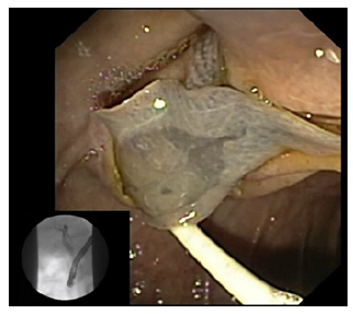
The authors report 3 cases of different patients with biliary parasitosis, diagnosed, and treated by endoscopic retrograde cholangiopancreatography (ERCP) (online suppl. video). All patients denied relevant risk factors for parasitosis, namely environmental, hygiene, or dietary factors. The first 2 cases illustrate biliary colonization by Fasciola hepatica. One of these patients presented with obstructive jaundice and the other with recurrent pancreatitis (two previous acute pancreatitis with mild dilation of extrahepatic bile ducts with biliary sludge in the common bile duct, described on emergency ultrasound). In the first case, abdominal ultrasound was suggestive of biliary parasites. In both patients, cholangiography performed during ERCP revealed multiple small filling defects with elliptic and sigmoidal shapes in the main bile duct with common bile duct dilation. After clearance of the bile ducts using an extraction balloon, the diagnosis was confirmed to be biliary parasitosis due to Fasciola hepatica. In the second case, the parasite was retrieved alive (shown in Fig. 1). Each patient received complementary treatment with triclabendazole the day after the endoscopic procedure at a dose of 10 mg/kg orally twice daily for 2 days.

Fig. 1 ERCP revealing removal of Fasciola hepatica through the papilla. 

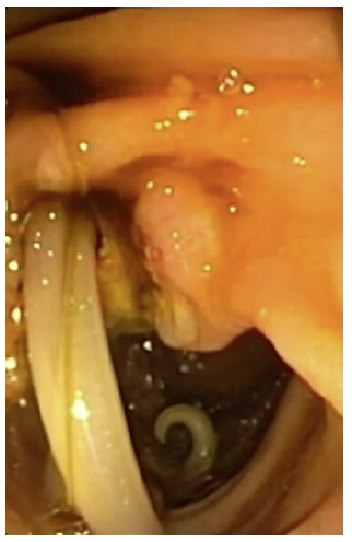
The third case corresponds to a patient that presented with acute cholangitis. During ERCP, cholangiography revealed many elongated irregular filling defects and dilation of the common bile duct without intrahepatic duct dilation. After performing sphincterotomy, the bile duct was cleared using a Dormia basket and a balloon extractor, allowing the removal of Ascaris lumbricoides already without motility (shown in Fig. 2). Endoscopic treatment was also supplemented with albendazole 400 mg orally as a single dose the day after the procedure. All patients had significant clinical improvement shortly after ERCP and have been discharged asymptomatic after 3 days.

Fig. 2 ERCP revealing removal of Ascaris lumbricoides through the papilla. 

In Portugal, as in other developed countries, biliary parasitosis are rare diseases. However, the incidence of these infestations appears to be increasing in line with the increase in tourism and immigration. Diagnosis is usually supported by ultrasound, computed tomography, magnetic resonance imaging (MRI), or endoscopic ultrasound [1]. Although ERCP is mainly used for therapeutic aspects, in rare cases ERCP is the key to the diagnosis [2]. We intend to illustrate the typical findings of parasitosis in cholangiography and duodenoscopy and demonstrate the usefulness of ERCP in the diagnosis and treatment of biliary obstructions due to biliary parasitosis.

Rerences

1. Garcia-Espinoza JA, Vásquez-Ciriaco S, Doña-Jaimes R, Aragon-Soto R, Velazco-Budar C, López-Martínez E. Parasitosis in the bile duct, report of 3 cases and literature review. Rev Médica del Hosp Gen México. 2018 Apr;81: 18-23. [ Links ]

2. Sezgin O, Altintas E, Tombak A, Ucbilek E. Fasciola hepatica-induced acute pancreatitis: report of two cases and review of the literature. Turkish J Gastroenterol. 2010 Jun 1;21(2):183-7. [ Links ]

1Statement of Ethics The study has been granted an exemption from requiring ethics approval in July 2022 (ULSAM Ethics Committee). Subjects have given their written informed consent to participate and publish their case and any accompanying images. Subjects have given their written informed consent to publish their case and any accompanying images.

3Funding Sources The authors have no funding sources to declare.

5Data Availability Statement All data generated or analyzed during this study are included in this article. Further inquiries can be directed to the corresponding author.

Received: September 30, 2022; Accepted: November 27, 2022

Correspondence to: Ana Catarina Carvalho, anacatarina54@gmail.com

Conflict of Interest Statement The authors have no conflicts of interest to declare.

Author Contributions Ana Catarina Carvalho: drafted the manuscript and performed video edition. Marta Moreira: acquired data and performed image edition. Luís Lopes: was responsible for conception and performance of the endoscopic procedure and critically revised the report. All authors read and approved the final version of the manuscript.

Creative Commons License This is an open-access article distributed under the terms of the Creative Commons Attribution License