


 Inglês (pdf)
 Artigo em XML
 Referências do artigo
 Enviar este artigo por email
 Citado por SciELO
  Similares em
SciELO
Similares em
SciELO 
 Permalink
A 65-year-old male patient was admitted to hospital with alcoholic steatohepatitis. He underwent esophagogastroduodenoscopy to assess for upper gastrointestinal (GI) bleeding due to hemoglobin levels of 7.4 g/dL. While neither an active/potential bleeding source nor esophagogastric varices were to be identified, a tiny, nipple-like, whitish lesion, with an estimated diameter of 1.5 mm, was detected in the distal esophagus 1 cm above the Z-line (Fig. 1a). To improve visualization and operability during intended forceps removal, we switched to cap-fitted endoscopy with a long, small-diameter cap (Fig. 1b). The lesion was grasped under visual control (Fig. 1c), resulting in a macroscopic complete one-piece resection (Fig. 1d). Pathologic evaluation of the targeted biopsy indicated squamous epithelium with hyperorthokeratosis, and a prominent granular layer, resembling the epidermis of the skin (Fig. 1e, f). Overall, the lesion was characterized as epidermoid metaplasia without signs of dysplasia.
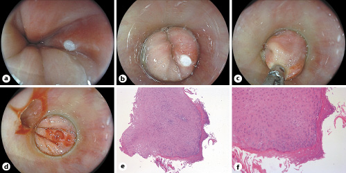
Fig. 1: a Standard endoscopy identified a compact-appearing, flat, elevated, whitish lesion of about 1.5 mm in size in the distal esophagus 1 cm above the Z-line. b Cap-fitted endoscopy likewise confirmed these findings and proved instrumental in the stabilization of the lesion for targeted forceps removal (c). d Post-biopsy assessment indicating macroscopic complete en bloc removal of the lesion. e, f Pathologic findings were significant for hyperorthokeratosis and a prominent granular layer without dysplasia, reminiscent of the epidermis of the skin (“epidermoid metaplasia”). HE. e ×5. f ×10.
Epidermoid metaplasia of the esophagus is rare in endoscopy practice, with only 2 cases (0.2%) being reported in 1,048 consecutive esophageal biopsies in the pathology literature [1]. Its distinct pathogenesis has still to be delineated, but chronic irritation, by, for example, alcohol and/or tobacco has been assumed to be contributory factors. Of note, epidermoid metaplasia has to be considered a preneoplastic lesion, such that exclusion of dysplasia requires meticulous endoscopic assessment of the whole esophageal mucosa, and, although not systematically studied and/or in a stricter sense evidence-based, surveillance endoscopies from time to time [2, 3]. Indeed, we have advised our patient to undergo a repeat upper endoscopy in 12 months. While most reports describe wide-spread lesions, to the best of our knowledge, this tiny focal lesion represents the smallest ever reported lesion. Albeit purely speculative, the emergence of this lesion could be attributable to defective healing complicating focal esophageal injury, e.g., esophagogastric reflux.

