SciELO - Scientific Electronic Library Online

 
vol.28 número5Repermeabilização esofágica anterógrada por assistência retrógrada: Abrindo novos caminhosSíndrome de ball-valve, uma apresentação incomum de GIST índice de autoresíndice de assuntosPesquisa de artigos
Home Pagelista alfabética de periódicos  

Serviços Personalizados

Journal

Artigo

Indicadores

Links relacionados

  • Não possue artigos similaresSimilares em SciELO

Compartilhar


GE-Portuguese Journal of Gastroenterology

versão impressa ISSN 2341-4545versão On-line ISSN 2387-1954

GE Port J Gastroenterol vol.28 no.5 Lisboa out. 2021  Epub 20-Mar-2022

https://doi.org/10.1159/000512360 

Endoscopic Snapshot

Underwater Duodenal ESD of a Large Adenoma Using the Pocket-Creation Method

“Underwater ESD” de um adenoma duodenal utilizando o “pocket-creation method”

João Santos-Antunesa  b  c 

Rui Moraisa 

Margarida Marquesa 

Guilherme Macedoa 

aGastroenterology Department, Faculty of Medicine, Centro Hospitalar S. João, Porto, Portugal;

bi3S-Instituto de Investigação e Inovação em Saúde, Universidade do Porto, Porto, Portugal;

cIPATIMUP - Institute of Molecular Pathology and Immunology, University of Porto, Porto, Portugal


Keywords Endoscopic submucosal dissection; Duodenal adenoma; Underwater ESD; Pocket-creation technique; Duodenal ESD

Palavras Chave Dissecção endoscópica da submucosa; Adenoma duodenal; ESD duodenal; Underwater ESD; Pocket-creation technique

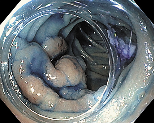
A 64-year-old woman, without relevant medical history, was referred to our department due to a 5-cm adenoma in the distal part of the second portion of the duodenum (Fig. 1). Due to the instability of the endoscope, the lesion size and localization, a complete resection by piecemeal-EMR was unlikely. Therefore, ESD was offered.

Fig. 1 Duodenal adenoma. 

We used hydroxyethylamide with methylene blue for submucosal injection, and dual- and IT-nano knives (Olympus, Tokyo) for submucosal dissection. We started in the oral side by a small incision in the mucosa and then performed underwater ESD using a total of nearly 1 L of saline, creating a pocket beneath the lesion, with the uneventful en bloc resection of the entire lesion.

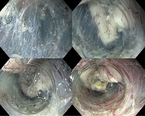
One of the major challenges in duodenal ESD is the absence of the contraction of the lesion after mucosal incision, which prevents an easy access to submucosa; at the same time, the muscle layer is very thin and the submucosal injection does not last long. Underwater technique allowed us to have an easier access to the submucosa, by pushing the mucosal flap away from the scope, and providing a clean visualization during the entire procedure. Once in the submucosa, pocket technique [1] allowed us to have a better scope stabilization throughout the procedure (Fig. 2); after dissecting the submucosa, we finished by cutting the remaining mucosa circularly.

Fig. 2 Underwater ESD and pocket-creation technique. 

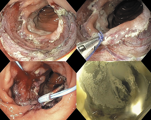
Besides the high-risk of adverse events during the procedure, duodenal ESD has a high risk of late adverse events, due to the exposure of the muscular layer to biliary and pancreatic juices. Therefore, we attempted to close the defect by the string-clip suturing method [2]. First, a clip with a string was passed into the endoscopic channel and displaced in the anal side of the ulcer. Then, a second clip was hooked in the string and displaced in the opposite (oral) side, and the two clips were gathered by pulling the string. After approaching the two margins, we tried to achieve the complete closure. Unfortunately, we were only able to close half of the defect, due to the poor mobility of the duodenum. In order to offer some protection to the muscle layer, we decided to prophylactically apply hemospray in the open area of the ulcer (Fig. 3).

Fig. 3 ESD defect; string-clip suturing method; hemospray. 

The lesion measured 60 × 40 mm and the procedure took 150 min. The patient was admitted to the hospital and stayed there for 1 week to address possible late adverse events related to the inability of complete closure of the defect, with double-dose proton pump inhibitor, that maintained for 8 weeks, and a stepwise progression to a normal diet. No late adverse events were observed.

Histological evaluation showed a high-grade dysplastic lesion, with free margins. Follow-up endoscopy showed a scar with some regenerative tissue; biopsies confirmed the absence of dysplasia.

Duodenal submucosal dissection has, in the West, limited indications due to the high rate of adverse events [3]. No previous Portuguese ESD series included underwater duodenal ESD [4-7]. However, when EMR is not possible, and considering the potentially aggressive surgeries, this can be attempted by experienced endoscopists in ESD.

References

1 Miura Y, Shinozaki S, Hayashi Y, Sakamoto H, Lefor AK, Yamamoto H. Duodenal endoscopic submucosal dissection is feasible using the pocket-creation method. Endoscopy. 2017 Jan;49(1):8-14. [ Links ]

2 Nishizawa T, Akimoto T, Uraoka T, Mitsunaga Y, Maehata T, Ochiai Y, et al. Endoscopic string clip suturing method: a prospective pilot study (with video). Gastrointest Endosc. 2018 Apr;87(4):1074-8. [ Links ]

3 Pimentel-Nunes P, Dinis-Ribeiro M, Ponchon T, Repici A, Vieth M, De Ceglie A, et al. Endoscopic submucosal dissection: European Society of Gastrointestinal Endoscopy (ESGE) Guideline. Endoscopy. 2015 Sep;47(9):829-54. [ Links ]

4 Santos-Antunes J, Baldaque-Silva F, Marques M, Lopes J, Carneiro F, Macedo G. Real-life evaluation of the safety, efficacy and therapeutic outcomes of endoscopic submucosal dissection in a Western tertiary centre. United European Gastroenterol J. 2018 Jun;6(5):702-9. [ Links ]

5 Costa RS, Ferreira A, Leal T, Costa D, Rolanda C, Gonçalves R. Endoscopic submucosal dissection for the treatment of superficial epithelial gastric neoplasia in a Portuguese Center. GE Port J Gastroenterol. 2019 Mar;26(2):90-8. [ Links ]

6 Rodrigues J, Carmo J, Carvalho L, Barreiro P, Chagas C. Endoscopic submucosal dissection for gastrointestinal superficial lesions: initial experience in a single Portuguese center. GE Port J Gastroenterol. 2015 Jul;22(5):190-7. [ Links ]

7 Santos-Antunes J, Marques M, Macedo G. Endoscopic Submucosal Dissection: experience in Portugal. GE Port J Gastroenterol. 2020 Jan;27(1):71-2. [ Links ]

Statement of Ethics The patient gave informed consent for the ESD procedure

Funding Source No funding was received

Received: August 08, 2020; Accepted: September 17, 2020

Corresponding author João Santos-Antunes Gastroenterology Department, Centro Hospitalar S. João Al. Prof. Hernani Monteiro PT-4200-319 Porto (Portugal) joao.claudio.antunes@gmail.com

Conflict of Interest Statement

The authors have no conflicts of interest to declare

Author Contributions

João Santos-Antunes performed the ESD and wrote the manuscript. Rui Morais, Margarida Marques and Guilherme Macedo provided critical revision of the manuscript

Creative Commons License This is an open-access article distributed under the terms of the Creative Commons Attribution License