


Serviços Personalizados
Journal
Artigo
 Inglês (pdf)
 Artigo em XML
 Referências do artigo
 Enviar este artigo por email
Indicadores
-
 Citado por SciELO
 -
 Acessos
Links relacionados
-
 Similares em
SciELO
Similares em
SciELO
Compartilhar

 Permalink
GE-Portuguese Journal of Gastroenterology
versão impressa ISSN 2341-4545
GE Port J Gastroenterol vol.27 no.2 Lisboa abr. 2020
https://doi.org/10.1159/000501399
IMAGES IN GASTROENTEROLOGY AND HEPATOLOGY
Sweets Syndrome as Manifestation of Active Ulcerative Colitis
A síndrome de Sweet como manifestação extra-intestinal de colite ulcerosa ativa
Mario Bento-Miranda, David N. Perdigoto, Sofia Mendes, Francisco Portela
Serviço de Gastrenterologia, Centro Hospitalar e Universitário de Coimbra, Coimbra, Portugal
* Corresponding author.
Keywords: Ulcerative colitis, Inflammatory bowel disease, Extraintestinal manifestations, Golimumab, Infliximab
Palavras-Chave: Colite ulcerosa, Doença inflamatória intestinal, Manifestações extra-intestinais, Golimumab, Infliximab
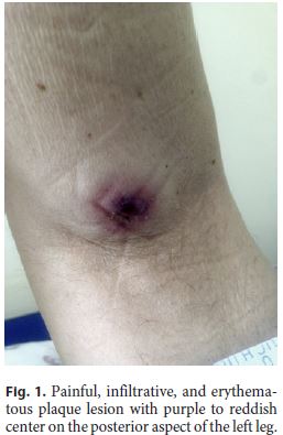
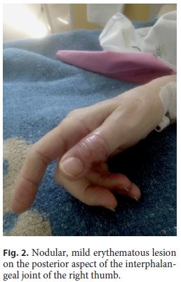
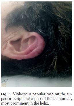
A 63-year-old woman with an extensive ulcerative colitis (UC) diagnosed over a decade ago presented with a history of abrupt onset of fever and erythematous nodular skin lesions, as well as increased bloody diarrhea and osteoarticular pain. She had been treated with mesalazine, azathioprine (suspended due to side effects), and infliximab (IFX). Physical examination showed the presence of erythematous skin lesions (Fig. 1, Fig. 2, Fig. 3). Blood analysis revealed elevated inflammatory markers, low circulating IFX levels, and significantly high anti-IFX antibodies. We performed a colonoscopy which evidenced signs compatible with active severe UC (Mayo endoscopic subscore: 3). Consequently, the patient was started on systemic corticotherapy.
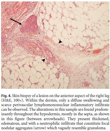
The skin lesions were biopsied (Fig. 4), allowing to establish the diagnosis of Sweets syndrome according to the revised criteria: abrupt onset of tender or painful erythematous plaques plus neutrophilic dermal infiltrate without leukocytoclastic vasculitis. Since a predominantly deep inflammatory infiltrate of hypodermis was acknowledged, this case was classified as a subcutaneous variant [1]. The patient also displayed variable features (minor criteria), such as fever and elevated inflammatory markers, which corroborates the diagnosis. Following this finding, we added a corticosteroid topical formulation for the skin lesions. Additionally, IFX was replaced by golimumab. The patient showed progressive improvement of both skin lesions and gastrointestinal symptoms, without relapsing. After discharge, methotrexate was added to the therapeutic plan.
Four months later, the patient was asymptomatic and with normalized inflammatory markers. The patient was kept on a high golimumab dose (100 mg in a patient with a body weight of 59 kg) due to the severity of the flair. We chose the 100-mg dose despite < 80 kg of body weight due to recent data pointing to lower trough levels in 50-mg non-responders, and methotrexate because the patient presents an increased risk of immunogenicity to the second anti-TNF. The treatment was discussed with the patient, and she was aware that full scientific support was lacking for both decisions [2].
Sweets syndrome is a rare extraintestinal manifestation of UC. This condition is an acute febrile neutrophilic dermatosis, characterized by a sudden reactive onset of painful erythematous skin papules and nodules that may coalesce into plaques, and is usually accompanied by fever and rising inflammatory markers (such as neutrophilia) [3].
In most cases, it is a neutrophil-mediated idiopathic hypersensitivity reaction. However, it may be associated with various factors, such as malignancies, infection, drugs, vaccination or, as in UC, inflammatory processes. Histologically, it translates as a dense neutrophilic infiltrate in the reticular dermis. One of the features that allows its differentiation from the other types of dermatosis is the absence of signs of leukocytoclastic vasculitis [1].
Treating the underlying condition is usually enough to attenuate or completely heal the cutaneous lesions, in most cases without scarring, although systemic, high-dose topic or intralesional corticosteroids may be necessary in more severe presentations. Up to one third of the patients have recurrent lesions [4].
References
1 Nofal A, Abdelmaksoud A, Amer H, Nofal E, Yosef A, Gharib K, et al. Sweets syndrome: diagnostic criteria revisited. J Dtsch Dermatol Ges. 2017 Nov;15(11):1081–8.
2 Philip G, Cornillie F, Adedokun JO, Melsheimer R, Rutgeerts P, Colombel JF, et al. Early dose optimization of golimumab in nonresponders to induction treatment for ulcerative colitis is effective and supported by pharmacokinetic data. J Crohns Colitis. 2019 Mar;jjz052. [ Links ]
3 Sweet RD. AN ACUTE FEBRILE NEUTROPHILIC DERMATOSIS. Br J Dermatol. 1964 Aug-Sep;76(8-9):349–56.
4 Hindryckx P, Novak G, Costanzo A, Danese S. Disease-related and drug-induced skin manifestations in inflammatory bowel disease. Expert Rev Gastroenterol Hepatol. 2017 Mar;11(3):203–14.
Statement of Ethics
I, the corresponding author, testify on behalf of all the co-authors that the subject mentioned gave her written informed consente to publish this case.
Disclosure Statement
F. Portela reports receiving lecture fees or serving as a board member from AbbVie, Celgene, Dr. Falk Pharma, Ferring Pharmaceuticals, Janssen Pharmaceuticals, Pfizer, Schering-Plough, and Takeda Pharmaceuticals. M. Bento-Miranda, D.N. Perdigoto, and S. Mendes have no conflicts of interest to declare.
* Corresponding author.
Dr. Mario Bento-Miranda
Serviço de Gastrenterologia
Centro Hospitalar e Universitário de Coimbra
Rua Virgílio Correia n° 70 3° direito, PT–3000-413 Coimbra (Portugal)
E-Mail mariobentomiranda@hotmail.com
Received: February 26, 2019; Accepted after revision: May 11, 2019
Acknowledgments
We thank Dr. José C. Cardoso for allowing us to use the figures of the skin biopsy.

