SciELO - Scientific Electronic Library Online

 
vol.10 issue3Cavernous Sinus Syndrome: An Unusual DiagnosisBeyond a Community-Acquired Pneumonia: Tracheoesophageal Fistula author indexsubject indexarticles search
Home Pagealphabetic serial listing  

Services on Demand

Journal

Article

Indicators

Related links

  • Have no similar articlesSimilars in SciELO

Share


Gazeta Médica

Print version ISSN 2183-8135On-line version ISSN 2184-0628

Gaz Med vol.10 no.3 Queluz Sept. 2023  Epub Sep 30, 2023

https://doi.org/10.29315/gm.v1i1.714 

Imagens Médicas

A Rare Entity with an Impressive Appearance

Uma Entidade Rara com uma Aparência Impressionante

1. Department of Pediatrics, Hospital de Braga, Braga, Portugal.

2. Department of Neonatology, Hospital de Braga, Braga, Portugal.

3. Department of Pediatric Surgery, Hospital de Braga, Braga, Portugal.


Keywords: Hemangioma/diagnosis; Infant, Newborn; Neoplasms, Vascular Tissue

Palavras-chave: Hemangioma/diagnóstico; Neoplasias de Tecido Vascular; Recém-Nascido

Introduction

Congenital hemangiomas (CH) are rare, benign vascular tumors typically located in the head, neck or limbs. Unlike infantile hemangiomas, they are present at birth and have their proliferative phase in utero.1,2 The three types of CH include the rapidly involuting (RICH), that involutes completely/nearly completely at 14 months; the noninvoluting (NICH), that does not resolve spontaneously and the partially involuting (PICH).1-3 Diagnosis is based on history and physical examination. Ultrasonography, MRI, arteriography and biopsy may be helpful when the diagnosis is uncertain.1,4 Complications include ulceration, bleeding, scarring, atrophy, thrombocytopenia and high-output cardiac failure.1,3 Treatment depends on tumor size, complications and tendency to spontaneous involution.1,4

Case Report

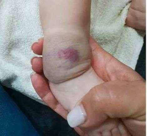
We report the case of a full-term female neonate, born by eutocic delivery, with a birth weight of 3175 g. Pregnancy was uneventful, with normal ultrasounds. Physical examination at birth revealed a raised, violaceous, soft-tissue mass at the right lateral malleolus, warm to touch, with prominent red small vesicles, measuring 3x4 cm (Fig. 1) and inguinal adenopathies in the ipsilateral limb. Doppler ultrasound revealed a well-defined hyperechogenic nodular image, with homogeneous texture and diffuse vascularity. There were no internal calcifications, and it did not extend to the profound plans. Magnetic resonance imaging (MRI) was performed for better characterization, revealing a benign tumoral vascular lesion (hemangioma or venous malformation), making the possibility of angiosarcoma or kaposiform hemangioendothelioma unlikely. Electrocardiogram, echocardiogram and transfontanelar ultrasound were normal. At four months, she has normal growth and development, and the lesion is undergoing rapid involution (discoloration and reduction in volume), with areas of skin redundancy, atrophy and changes in texture with persistent scattered veins and telangiectasias (Fig. 2).

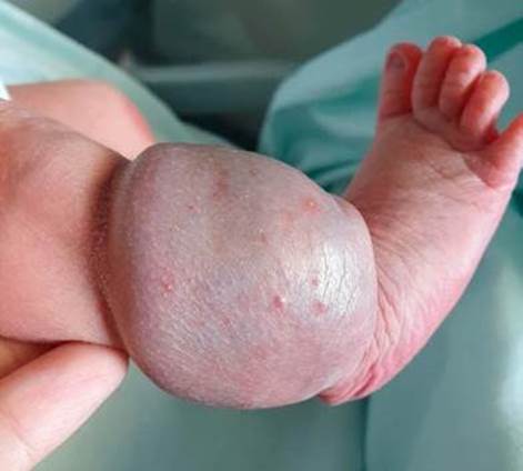
Figure 1: At birth: Soft-tissue mass, raised, violaceous, warm to touch, with prominent red small vesicles, measuring 3x4 cm and inguinal adenopathies in the ipsilateral member. 

Figure 2: At four-month-old: lesion with discoloration and reduction in the volume, with areas of skin redundancy, atrophy and changes in texture. 

Conclusion

Although RICH have an impressive appearance, they are benign and most of them improve without treatment. Clinicians must recognize and differentiate CH from malignant entities.

References

1. Fieden I, Adams D. Congenital hemangiomas: Rapidly involuting congenital hemangioma (RICH), noninvoluting congenital hemangioma (NICH), and partially involuting congenital hemangioma (PICH). UpToDate. [accessed January 2022] Available from: Available from: https://www.uptodate.com/contents/congenital-hemangiomas-rapidly-involuting-congenital-hemangioma-rich-noninvoluting-congenital-hemangioma-nich-and-partially-involuting-congenital-hemangioma-pich . [ Links ]

2. Krol A, MacArther C. Congenital hemangiomas. Arch Facial Plast Surg. 2005;7:307-11. doi: 10.1001/archfaci.7.5.307. [ Links ]

3. Boon L, Enjolras O, Mulliken J. Congenital hemangioma: Evidence of accelerated involution. J Pediatr. 1996;128:329-35. doi: 10.1016/s0022-3476(96)70276-7. [ Links ]

4. Browning J, Metry D. Rapidly involuting congenital hemangioma: case report and review of the literature. Dermatol Online J. 2008;14:11. [ Links ]

Declaração de Contribuição/Contributorship Statement

MS e NS: Pesquisa e escrita do artigo

ASR, ARR e SBM: Pesquisa e revisão do artigo

MS and NS: Research and article writing

ASR, ARR and SBM: Research and article review

Responsabilidades Éticas

Conflitos de Interesse: Os autores declaram a inexistência de conflitos de interesse na realização do presente trabalho.

Fontes de Financiamento: Não existiram fontes externas de financiamento para a realização deste artigo.

Confidencialidade dos Dados: Os autores declaram ter seguido os protocolos da sua instituição acerca da publicação dos dados de doentes.

Consentimento: Consentimento do doente para publicação obtido.

Proveniência e Revisão por Pares: Não comissionado; revisão externa por pares.

Ethical Disclosures

Conflicts of Interest: The authors have no conflicts of interest to declare.

Financing Support: This work has not received any contribution, grant or scholarship.

Confidentiality of Data: The authors declare that they have followed the protocols of their work center on the publication of data from patients.

Patient Consent: Consent for publication was obtained.

Provenance and Peer Review: Not commissioned; externally peer reviewed.

Received: March 07, 2023; Accepted: June 16, 2023; preprint: July 07, 2023; Published: September 30, 2023

Autor Correspondente/Corresponding Author: Mariana Santos [marianamltsantos@gmail.com] ORCID iD: 0000-0002-9612-6681

Creative Commons License This is an open-access article distributed under the terms of the Creative Commons Attribution License