Imagens Médicas
Cavernous Sinus Syndrome: An Unusual Diagnosis
Síndrome do Seio Cavernoso: Um diagnóstico Pouco Frequente
1. Internal Medicine Department, Hospital Distrital da Figueira da Foz, Figueira da Foz, Portugal.
2. Internal Medicine Department, Centro Hospitalar Médio Tejo, Tomar, Portugal.
Abstract
Cavernous sinus syndrome is characterized by ophthalmoplegia, diplopia, facial hypoesthesia, Horner syndrome and proptosis.
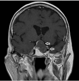
The authors present a clinical case of an 89-year-old woman presented to the emergency department with complaints of vomiting, severe left frontotemporal headache, photophobia, left ptosis and diplopia with less than 24 hours of evolution. Neurological examination revealed ptosis and limitation of all eye movements in the left eye. Cranioencephalic computed tomography and magnetic resonance imaging showed an expansive lesion with expression in the suprasellar cistern and in the left cavernous sinus, suggestive of a macroadenoma with signs of hemorrhagic transformation.
Cavernous sinus syndrome has several etiologies, as cerebrovascular diseases, infectious diseases, and trauma. In addition to the clinical history and physical examination, imaging tests are essential to determine the etiology behind this syndrome.
Keywords: Adenoma; Cavernous Sinus; Pituitary Neoplasms
Resumo
A síndrome do seio cavernoso caracteriza-se pela presença de oftalmoplegia, diplopia, hipostesia facial, síndrome de Horner e proptose. Os autores apresentam o caso clínico de uma mulher, 89 anos, que se apresentou no Serviço de Urgência com queixas de vómitos, cefaleia frontotemporal esquerda, fotocopia, ptose esquerda e diplopia com cerca de 24 horas de evolução. O exame neurológico revelou a presença de ptose e alteração nos movimentos oculares ao nível do olho esquerdo. A tomografia computorizada e ressonância magnética crânio-encefálica revelaram a presença de lesão expansiva na região supraselar e seio cavernoso esquerdo, sugestiva de macroadenoma com sinais de transformação hemorrágica. A síndrome do seio cavernoso relaciona-se com diversas etiologias, tais como, doenças cerebrovasculares, infecciosas e trauma. Para além da história clínica e exame objetivo, os exames imagiológicos são essenciais para o diagnóstico e determinação da etiologia desta síndrome.
Palavras-chave: Adenoma; Neoplasias Pituitárias; Seio Cavernoso
An 89-year-old woman went to the emergency department with complaints of vomiting, severe left frontotemporal headache, photophobia, left ptosis and diplopia with less than 24 hours of evolution.
Neurological examination revealed ptosis and limitation of all eye movements in the left eye. She also presented hypoesthesia in the territory of the maxillary branch of the left trigeminal nerve, with no other focal alterations.
Cranioencephalic computed tomography (CT) and magnetic resonance imaging (MRI) showed an expansive lesion with expression in the suprasellar cistern and in the left cavernous sinus, suggestive of a macroadenoma with signs of hemorrhagic transformation.” (Fig. 1).
The diagnosis of cavernous sinus syndrome (paresis of left cranial nerves III, IV and V2) was made, resulting from a macroadenoma with cavernous sinus invasion. The patient underwent endoscopic transphenoidal resection of the macroadenoma, with favorable clinical and imaging course and was later discharged from the hospital.
The cavernous sinus syndrome (CSS) is a set of signs and symptoms, including ophthalmoplegia, diplopia, facial hypoesthesia, Horner syndrome and proptosis.1 Involvement of the III and IV cranial nerves can result in ophthalmoplegia and diplopia, while the involvement of the ophthalmic and maxillary divisions of the trigeminal nerve can result in altered sensitivity on the face.2 CSS have several etiologies, as cerebrovascular diseases, infectious diseases, and trauma.1
In addition to the clinical history and physical examination, imaging tests, especially MRI with a study aimed at cavernous sinus, are essential to determine the etiology behind this syndrome,3 as was the case with this patient.
References
1. Nambiar R, Nair SG. Cavernous sinus syndrome. Bayl Univ Med Cent Proc. 2017;30:455-456. doi: 10.1080/08998280.2017.11930227.
[ Links ]
2. Munawar K, Nayak G, Fatterpekar GM, Sen C, Zagzag D, Zan E, et al. Cavernous sinus lesions. Clin Imaging. 2020;68:71-89. doi: 10.1016/j.clinimag.2020.06.029.
[ Links ]
3. Bhatkar S, Mahesh KV, Sachdeva J, Goel A, Goyal MK, Takkar A, et al. Magnetic resonance imaging (MRI) versus computed tomographic scan (CT scan) of brain in evaluation of suspected cavernous sinus syndrome. Neuroradiol J. 2020;33:501-7. doi: 10.1177/1971400920970921.
[ Links ]


 Inglês (pdf)
 Artigo em XML
 Referências do artigo
 Enviar este artigo por email
 Citado por SciELO
  Similares em
SciELO
Similares em
SciELO 
 Permalink