SciELO - Scientific Electronic Library Online

 
vol.9 número3Neoformação Renal: Um Diagnóstico a ConsiderarSarcoma de Kaposi da Cavidade Oral índice de autoresíndice de materiabúsqueda de artículos
Home Pagelista alfabética de revistas  

Servicios Personalizados

Revista

Articulo

Indicadores

Links relacionados

  • No hay articulos similaresSimilares en SciELO

Compartir


Gazeta Médica

versión impresa ISSN 2183-8135versión On-line ISSN 2184-0628

Gaz Med vol.9 no.3 Queluz set. 2022  Epub 30-Sep-2022

https://doi.org/10.29315/gm.v1i1.594 

Casos Clínicos

“Pseudo-Geyser Sign” as the First Presentation of Septic Arthritis of the Shoulder

“Pseudo-Geyser Sign” como Primeira Apresentação de Artrite Sética do Ombro

Madalena Braga1 
http://orcid.org/0000-0002-3808-2705

Joana P. Sousa2 

João Torres3  4 

1. USF Arca D’Água, ACeS Porto Oriental, Porto, Portugal.

2. USF Faria Guimarães, ACeS Porto Oriental, Porto, Portugal.

3. Centro Hospitalar Universitário de S. João, Porto, Portugal.

4. Faculdade Medicina Universidade Porto, Porto, Portugal.


Abstract

Massive rotator cuff tears can lead to large cysts, extending from the subacromial space through the acromioclavicular joint into a subcutaneous cyst, with a typical “geyser sign” on magnetic resonance imaging (MRI). To our knowledge, no cases of transdeltoid pseudo-cyst as first manifestation of septic arthritis have ever been reported.

A 79-year-old female presented to the Emergency Department (ED) with a two-day evolution of a painful tumefaction over her right shoulder. She had been admitted to the ED 16 days before, after being bitten by a dog. Tumefaction drainage revealed a total cell count of 50320 cells/mL. MRI revealed glenohumeral joint cavity’s fluid “decompression” through the deltoid. Septic arthritis was presumed as the cause of the pseudo-cyst, configuring a “pseudo-geyser sign” on MRI.

Septic arthritis is an orthopedic emergency, whose early diagnosis, ED referral and treatment are crucial.

Keywords: Arthritis, Infectious; Rotator Cuff; Shoulder Joint

Resumo

Ruturas maciças da coifa dos rotadores podem levar à formação de grandes quistos, que se estendem do espaço subacromial, através da articulação acromioclavicular, formando um quisto subcutâneo, com um “geyser sign” típico na ressonância magnética (RM).

Até onde sabemos, nenhum caso de pseudo-quisto transdeltoide como primeira manifestação de artrite séptica foi relatado. Uma mulher de 79 anos recorreu ao Serviço de Urgência (SU) com uma tumefação dolorosa no ombro direito com dois dias de evolução. Ela tinha recorrido ao SU 16 dias antes, após ter sido mordida por um cão. A drenagem de tumefação revelou uma contagem total de células de 50320 células/mL. A RM revelou “descompressão” do fluido da cavidade articular glenoumeral através do deltoide. A artrite sética foi presumida como a causa do pseudoquisto, configurando um “pseudo-geyser sign” na RM.

A artrite sética é uma emergência ortopédica, cujo diagnóstico precoce, encaminhamento ao SU e tratamento são cruciais.

Palavras-chave: Artrite Infeciosa; Articulação do Ombro; Coifa dos Rotadores

Introduction

Several case reports have previously shown that massive rotator cuff tears can lead to large cysts extending from the subacromial space through the acromioclavicular joint into a subcutaneous cyst, with a characteristic “geyser sign” on imaging.1-6 One single and recent study has reported extension through the deltoid muscle.7 However, to our knowledge, no cases of transdeltoid pseudo-cyst as the first manifestation of septic arthritis have ever been reported.

Case Report

A 79-year-old female, with a clinical history of arterial hypertension and dyslipidemia, under telmisartan + hydrochlorothiazide, 80 mg + 25 mg, and simvastatin, 20 mg, both once daily, presented to the Emergency Department (ED) with a two-day evolution of a painful tumefaction over her right shoulder (Fig. 1). She denied any history of recent trauma. During the physical examination, she showed limited passive and active range of motion. A mass could be palpated in the lateral side of her right shoulder, measuring about 5 cm, associated with local heat, redness, swelling and pain. The redness was extended to the armpit area and multiple adenopathies could be palpated in the axillary region, with 2 to 3 cm. The tympanic temperature was 37.5ºC. The medical records showed the patient had been admitted to the hospital 16 days before, after being bitten by a neighbor’s dog on her left leg; objective examination was then normal, and she was discharged medicated with oral antibiotic therapy (amoxicillin + clavulanic acid, 875 mg + 125 mg, every 12 hours, for 8 days) and appropriate wound dressings’ advice. Right shoulder ultrasound on admission was notable for a voluminous and heterogeneous, yet inconclusive, subcutaneous fluid collection. Puncture and drainage of the lesion were performed, resulting in 10 mL of a pasty serohematic fluid; cytological examination revealed a total cell count of 50320 cells/mL, with a predominance of polymorphonuclear leukocytes (92.7%). Blood tests were notable for an increase in C-reactive protein levels (34.3 mg/dL). Magnetic resonance imaging (MRI) of the right shoulder was performed, which revealed massive rupture of the rotator cuff tendons involving the entire supraspinatus and infraspinatus and the upper segment of the subscapularis muscle. Also, it was notable for blurring of the tendon of the long portion of the biceps and moderate effusion in the glenohumeral and acromioclavicular joint cavities, with fluid leakage into the subacromial/ subdeltoid bursa. The glenohumeral joint cavity demonstrated "decompression" to the subcutaneous cell tissue through a linear defect in the lateral aspect of the deltoid muscle body, where it presented as a collection measuring about 33 x 30 x 40 mm (Figs. 2 and 3). Blood and synovial fluid cultures were negative. The diagnosis of septic arthritis was presumed based on the clinical history and articular fluid cytological examination. The patient was initially proposed for surgical debridement and irrigation of the right shoulder, not only to improve the prognosis, but also to perform the culture of the synovial fluid collected during surgery and possibly target antibiotic therapy. However, given the clinical and analytical improvement of the patient condition during hospital stay, it was decided not to undergo surgical treatment.

Figure 1: Tumefaction presented by the patient in the emergency department. 

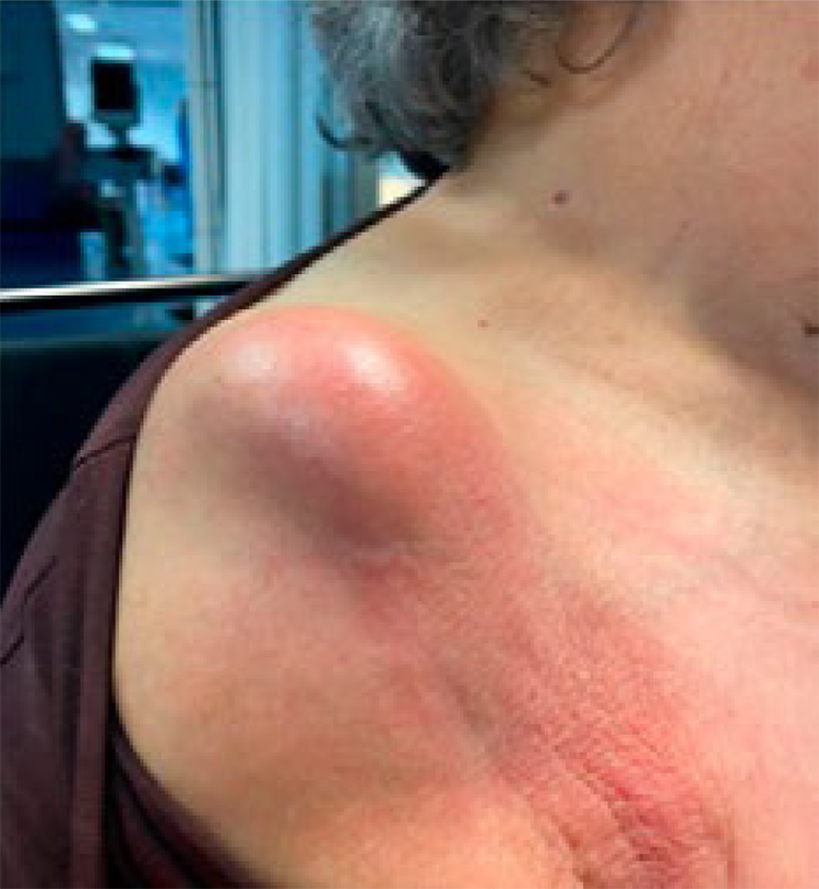
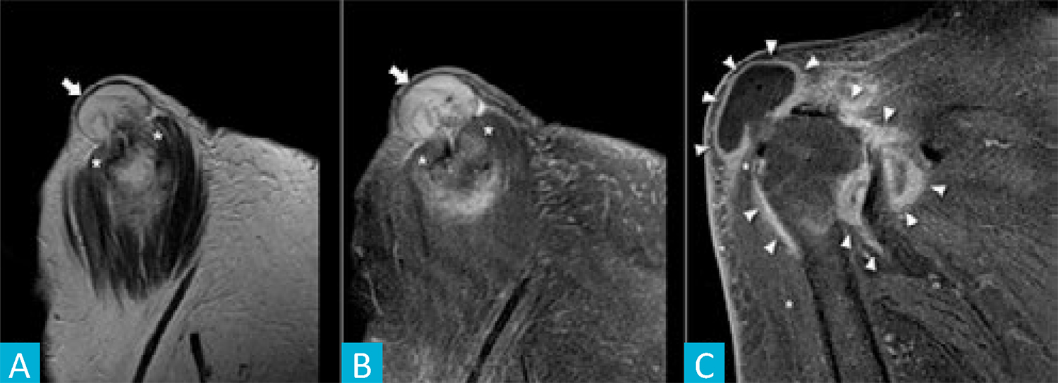
Figure 2: Pseudo-geyser. Sagital T2w weighted (A) and sagital T2w fat-saturated (B) MR images depicting the pseudo-geyser [arrow] protruding through a full thickness tear near the proximal insertion of the deltoid [asterisk]. Coronal T1w fat-saturated MR image after intravascular administration of gadolinium (C) showing the hyper-enhancement of the pseudo-geyser wall and the articular capsule, corresponding to the inflammatory/ infectious reaction. 

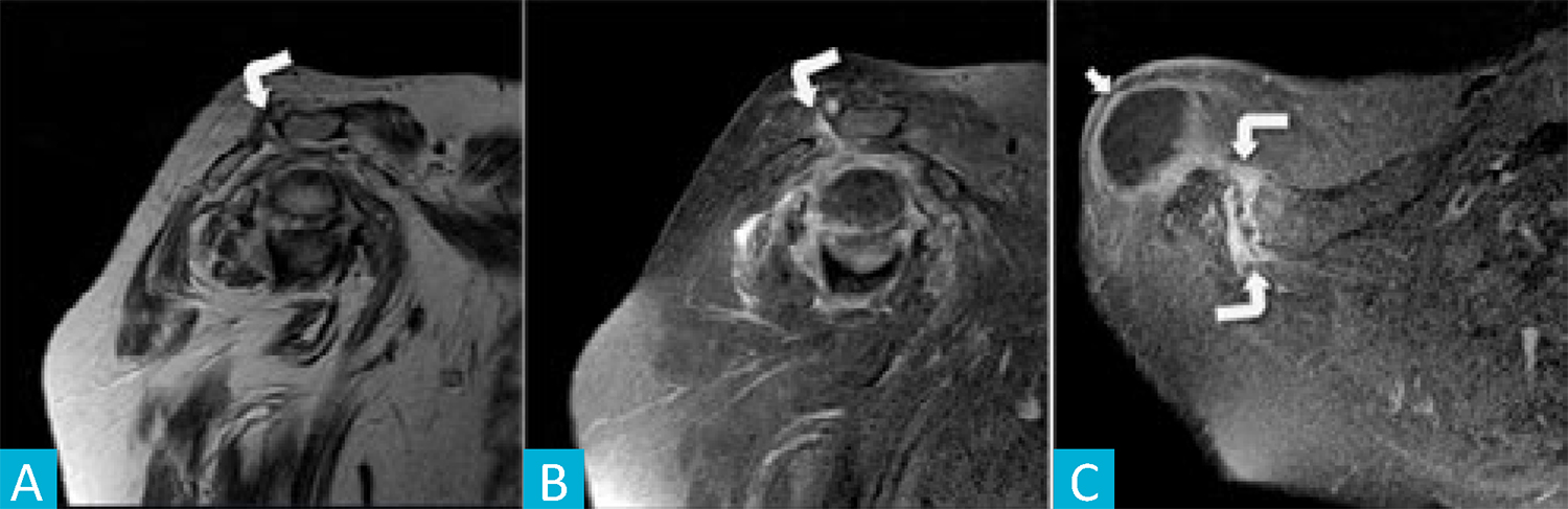
Figure 3: Pseudo-geyser. Sagital T1w (A), sagital T2w fat-saturated (B) and axial T1w fat-saturated after intravascular gadolinium (C) MR images depicting the acromioclavicular joint [curved arrow] with some degenerative changes, effusion and periarticular edema but with no relationship to the pseudo-geyser [arrow]. 

Conclusion

Septic arthritis, also known as infectious arthritis, represents a direct invasion of a joint space by various microorganisms, mainly bacteria,8 and the most common route of invasion of the joint is via the bloodstream.9 The MRI scan revealed a fluid-filled cyst extending from the subacromial space through the deltoid in the context of a long-standing massive rotator cuff tear, configuring a “pseudo-geyser sign” on imaging. Its growth and exuberant inflammatory signs were thought to be the result of an episode of septic arthritis in the context of apparent hematogenous spread due to the previous dog bite incident. Prior antibiotic exposure can justify not only the negative blood and synovial fluid cultures, but also the self-limited course of the disease. Though uncommon, septic arthritis is an orthopedic emergency that can cause significant joint damage, leading to increased morbidity and mortality. Clinicians should be aware of the signs and symptoms of septic arthritis, owing to the fact that the early diagnosis, prompt referral to the ED and treatment are crucial.4

References

1. DiFelice GS. The MRI geyser sign: acromioclavicular joint cysts in the setting of a chronic rotator cuff tear. Am J Orthop. 2011;40:E118-E121. [ Links ]

2. Craig EV. The acromioclavicular joint cyst. An unusual presentation of a rotator cuff tear. Clin Orthop Relat Res. 1986;202:189-92. [ Links ]

3. Lizaur UA, Marco Gomez L, Perez Aznar A, Cebrian Gomez R. Rotator cuff tear and acromioclavicular joint cyst. Acta Orthop Belg. 1995;61:144-6. [ Links ]

4. Gumina S. Acromioclavicular joint cyst associated with rotator cuff tear. A report of three cases. Clin Orthop Relat Res. 1993;294:111-3. [ Links ]

5. Selvi E, De Stefano R, Frati E, Manganelli S, Manca S, Marcolongo R. Rotator cuff tear associated with an acromioclavicular cyst in rheumatoid arthritis. Clin Rheumatol. 1998;17:170-1. doi: 10.1007/BF01452269. [ Links ]

6. Tshering Vogel DW, Steinbach LS, Hertel R, Bernhard J, Stauffer E, Anderson SE. Acromioclavicular joint cyst: nine cases of a pseudotumor of the shoulder. Skeletal Radiol. 2005;34:260-5. doi: 10.1007/s00256-004-0883-6. [ Links ]

7. Martin JR, Amini MH. Deltoid defect from a trans-deltoid synovial cyst in a patient undergoing reverse shoulder arthroplasty: a case report. JSESInt. 2021;5.1:138.doi: 10.1016/j.jseint.2020.10.001. [ Links ]

8. Roerdink RL, Huijbregts HJ, van Lieshout AW, Dietvorst M, van der Zwaard BC. The difference between native septic arthritis and prosthetic joint infections: A review of literature. J Orthop Surg. 2019;27:2309499019860468. doi: 10.1177/2309499019860468. [ Links ]

9. Rice PA. Gonococcal arthritis (disseminated gonococcal infection). Infect Dis Clin North Am. 2005;19:853-61. doi: 10.1016/j.idc.2005.07.003. [ Links ]

Presentations and Awards/Apresentações e Prémios

1The article was submitted for presentation at WONCA Europe Conference 2022 and is awaiting approval.

Authors Contribution/Contribuição Autoral

MB: Study preparation, design and writing

JPS: Study design and writing

JT: Mentoring, study design, writing

MB: Preparação, desenho e redação do estudo

JPS: Conceção e redação do estudo

JT: Mentoria, desenho e redação do estudo

Responsabilidades Éticas

Conflitos de Interesse: Os autores declaram a inexistência de conflitos de interesse na realização do presente trabalho.

Fontes de Financiamento: Não existiram fontes externas de financiamento para a realização deste artigo.

Confidencialidade dos Dados: Os autores declaram ter seguido os protocolos da sua instituição acerca da publicação dos dados de doentes.

Consentimento: Consentimento do doente para publicação obtido.

Proveniência e Revisão por Pares: Não comissionado; revisão externa por pares.

Ethical Disclosures

Conflicts of Interest: The authors have no conflicts of interest to declare.

Financing Support: This work has not received any contribution, grant or scholarship.

Confidentiality of Data: The authors declare that they have followed the protocols of their work center on the publication of data from patients.

Patient Consent: Consent for publication was obtained.

Provenance and Peer Review: Not commissioned; externally peer reviewed.

Received: May 02, 2022; Accepted: July 18, 2022; preprint: September 01, 2022; Published: September 30, 2022

*Corresponding Author/Autor Correspondente: Madalena Peixoto de Sousa Braga [madalena.braga3@gmail.com] Rua Prof. Fernando Magano, 140A, 4º esquerdo, 4250-541, Porto ORCID iD: 0000-0002-3808-2705

Creative Commons License This is an open-access article distributed under the terms of the Creative Commons Attribution License