SciELO - Scientific Electronic Library Online

 
vol.35 issue3Radiation Recall MyositisRetropubic Cartilaginous Cyst: A Rare Entity author indexsubject indexarticles search
Home Pagealphabetic serial listing  

Services on Demand

Journal

Article

Indicators

Related links

  • Have no similar articlesSimilars in SciELO

Share


Acta Radiológica Portuguesa

Print version ISSN 2183-1351

Acta Radiol Port vol.35 no.3 Lisboa Dec. 2023  Epub Dec 29, 2023

https://doi.org/10.25748/arp.27357 

Imagens de Interesse

Tenosynovitis of the Peroneal Tendons Due to Hypertrophy of the Peroneal Tubercle - Diagnosis Using Ultrasound and Magnetic Resonance Imaging

Tenossinovite dos Tendões Fibulares Devido a Hipertrofia do Tubérculo Fibular - Diagnóstico Utilizando a Ultrassonografia e a Ressonância Magnética

Mayara Oliveira da Silva1 
http://orcid.org/0000-0002-9882-3456

Márcio Luís Duarte2 
http://orcid.org/0000-0002-7874-9332

1Universidade Federal de São Paulo (UNIFESP) - Campus Baixada Santista, Santos-SP, Brasil

2Universidade de Ribeirão Preto (UNAERP) - Campus Guarujá, Guarujá-SP, Brasil


Abstract

The prevalence of peroneal tubercles ranges from 24% to 98.58% - this wide range can be attributed to the use of different criteria in its description. The incidence of the hypertrophied tubercle is 20.5-24% of the population. The portion of the peroneus longus tendon that is located between the fixed bony points of the peroneal tubercle and the cuboid may stretch unnaturally in the presence of a hypertrophied peroneal tubercle and, with sudden or repeated inversion movements, the tendon may rupture. The peroneus brevis tendon can collide between the tubercle and the lateral malleolus during the eversion-abduction movement of the foot, resulting in tenosynovitis, tendon laceration, and degeneration. We report peroneal tenosynovitis due to a hypertrophied peroneal tubercle induced by frictional peroneal tendons activity detected by ultrasound and MRI.

Keywords: Calcaneus; Tenosynovitis; Ultrasonography; Magnetic resonance imaging; Diagnosis.

Resumo

A prevalência de tubérculos fibulares varia de 24% a 98,58% - essa ampla variação pode ser atribuída ao uso de diferentes critérios na descrição do tubérculo fibular. A incidência do tubérculo hipertrofiado é de 20,5-24% da população. A porção do tendão fibular longo que está localizada entre os pontos ósseos fixos do tubérculo fibular e do cubóide pode se estirar de forma não natural na presença de um tubérculo fibular hipertrofiado e, com movimentos de inversão repentinos ou repetidos, o tendão pode romper. Raramente, o tubérculo hipertrofiado está associado a uma rotura do tendão do fibular curto. O tendão do fibular curto pode ser colidido entre o tubérculo e o maléolo lateral durante o movimento de eversão-abdução do pé, resultando em tenossinovite e degeneração do tendão. Relatamos uma tenossinovite dos fibulares decorrente de um tubérculo fibular hipertrofiado induzido pela atividade friccional dos tendões fibulares detectada pela ultrassonografia e pela ressonância magnética.

Palavras-chave: Calcâneo; Tenossinovite; Ultrassonografia; Imagem por ressonância magnética; Diagnóstico.

The usual size of the peroneal tubercle is 3 mm.1 The size of the critical peroneal tubercle - borderline to promote tendon injury - is 4.3 mm on magnetic resonance imaging (MRI).1

It is supposed that most cases are congenital (flat or paralytic foot, equinus foot, insertion of the peroneus quartus muscle), but some are acquired by previous trauma, altered weight support, and/or inflammatory diseases related to spasms of the peroneus longus tendon.2 The portion of the peroneus longus tendon that is located between the fixed bony points of the peroneal tubercle and the cuboid may stretch unnaturally in the presence of a hypertrophied peroneal tubercle and, with sudden or repeated inversion movements, the tendon may rupture.3 The peroneus brevis tendon can collide between the tubercle and the lateral malleolus during the eversion-abduction movement of the foot, resulting in tenosynovitis, tendon laceration, and degeneration.1

The first symptoms occur when the peroneal hypertrophic tuber causes chronic irritation of the tendons and tendon sheaths, with the peroneus longus tendon being more vulnerable to damage than the peroneus brevis tendon owing to its length, sharp change of direction, and subsequent location to the peroneal tubercle.2 The typical finding on physical examination is edema in the lateral region adjacent to the calcaneal bone, focal tenderness over the peroneal tendons, and close to the peroneal tubercle, in addition to a positive peroneal tendon resistance test.1

A peroneal tubercle can be identified by an axial view of the calcaneus or anteroposterior radiographs of the ankle, while the size of the peroneal tubercle can be assessed by multidetector computed tomography and three-dimensional volume rendering.2 Ultrasound and MRI are the diagnostic methods indicated for the assessment of coexisting conditions of the peroneal tendons and their differential diagnoses.2 Furthermore, MRI can detect the hypertrophied peroneal tubercle edema that is induced by frictional peroneal tendon activity.1 The differential diagnosis of lateral ankle pain consists of injury to the lateral ankle ligaments, subluxation/dislocation of the peroneal tendons, peroneal tenosynovitis, and osteochondral lesions of the talus.2,3

The initial treatment is usually conservative, including shoe modifications, padding, bracing, and immobilization.2 However, of the lanyard to the systematic review of Lohrer et al,1 only one case was successful, and in other cases, surgical interventions are indicated, consisting of the removal of the hypertrophic peroneal tubercle and repair of the peroneal tendons.2,3

We report a 42-year-old male with pain in the lateral region of the hindfoot. Refers to worsening when walking for a long time. On physical examination, he presents pain in the eversion of the ankle and foot dorsiflexion, without functional limitation. He does not present edema but presents pain during palpation in the lateral region of the calcaneal bone.

Ultrasound detected echotexture alteration in the peroneal brevis tendon and mild liquid effusion adjacent to the peroneal tubercle (Fig. 1). MRI detected hypertrophy (0.6 mm) and significant edema of the peroneal tubercle (Fig. 2) with peroneal tenosynovitis (Fig. 3) and mild liquid effusion around the region of the skin marker. Those findings are compatible with tenosynovitis due to a hypertrophied peroneal tubercle induced by frictional peroneal tendon activity. The patient was referred to the orthopedics department for scheduled surgery.

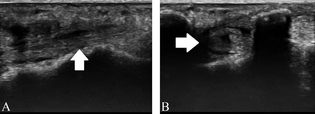
Figure 1: Ultrasound demonstrating tenosynovitis of the peroneal brevis (white arrow) in the sagittal section (A) and axial section (B). 

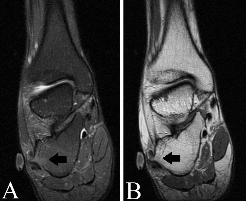
Figure 2: MRI in the coronal section of the right ankle, T2 FAT SAT weighted-image (A) and T2 weighted-image (B) demonstrating edema in the hypertrophied peroneal tubercle (black arrow). 

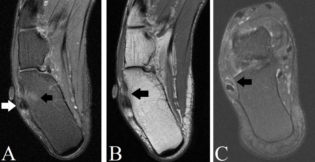
Figure 3: MRI in the axial section of the right ankle, T2 FAT SAT weighted-image (A) and DP weighted-image (B) demonstrating edema in the hypertrophied peroneal tubercle (black arrow) and tenosynovitis of the peroneal tendons (white arrow). In C, MRI in the oblique section of the right ankle in T2 FAT SAT weighted-image demonstrating marked tenosynovitis of the peroneal brevis tendon (black arrow). 

References

1. Lohrer H. Distal peroneus longus dislocation and pseudohypertrophy of the peroneal tubercle: a systematic review. J Foot Ankle Surg. 2019;58:969-73. [ Links ]

2. Celikyay F, Yuksekkaya R, Almus F, Bilgic E. Tenosynovitis of the peroneal tendons associated with a hypertrophic peroneal tubercle: radiography and MRI findings. BMJ Case Rep. 2014;2014:bcr2013200204. [ Links ]

3. Boya H, Pinar H. Stenosing tenosynovitis of the peroneus brevis tendon associated with hypertrophy of the peroneal tubercle. J Foot Ankle Surg . 2010;49:188-90. [ Links ]

Ethical Disclosures

Financing Support: This work has not received any contribution, grant or scholarship.

Received: June 09, 2022; Accepted: August 31, 2022

Address Márcio Luís Duarte, Universidade de Ribeirão Preto (UNAERP) - Campus Guarujá, Av. D. Pedro I, 3.300, Enseada, Guarujá-SP - Brasil. CEP: 11440-003, e-mail: marcioluisduarte@gmail.com

Conflicts of interest: The authors have no conflicts of interest to declare.

Confidentiality of data: The authors declare that they have followed the protocols of their work center on the publication of data from patients.

Protection of human and animal subjects: The authors declare that the procedures followed were in accordance with the regulations of the relevant clinical research ethics committee and with those of the Code of Ethics of the World Medical Association (Declaration of Helsinki).

© Author(s) (or their employer(s)) and ARP 2023. Re-use permitted under CC BY-NC. No commercial re-use.

Creative Commons License This is an open-access article distributed under the terms of the Creative Commons Attribution License