SciELO - Scientific Electronic Library Online

 
vol.35 issue2Vaginal Lithiasis - A Rare CaseAn Atypical Cause of Pain and Motor Weakness of the Left Forearm author indexsubject indexarticles search
Home Pagealphabetic serial listing  

Services on Demand

Journal

Article

Indicators

Related links

  • Have no similar articlesSimilars in SciELO

Share


Acta Radiológica Portuguesa

Print version ISSN 2183-1351

Acta Radiol Port vol.35 no.2 Lisboa Aug. 2023  Epub Aug 31, 2023

https://doi.org/10.25748/arp.27587 

Imagens de Interesse

Cervical Mass in an Infant

Massa Cervical num Lactente

1Pediatric Department, São Francisco Xavier Hospital, CHLO, Lisboa, Portugal

2Radiology Department, São Francisco Xavier Hospital, CHLO, Lisboa, Portugal


Abstract

Fibromatosis colli is the most severe presentation of congenital muscular torticollis and a relatively rare cause of neck swelling in neonates and infants. We present a case of a 6 weeks old male infant who was admitted to the emergency department with a mass in the left lateral-cervical region. Two soft tissue ultrasounds were performed showing multiple adenopathy and one week later a new ultrasound was performed by a radiologist experienced in pediatric imaging that was suggestive of fibromatosis colli. Ultrasound is the diagnosis imaging modality of choice if there is any doubt in the diagnosis. It shows focal or diffuse enlargement of the sternocleidomastoid muscle in a fusiform configuration.

Keywords: Cervical mass; Fibromatosis colli; Congenital torticollis.

Resumo

A Fibromatosis colli é a apresentação mais grave do torcicolo muscular congénito e uma causa rara de tumefação cervical em recém-nascidos e lactentes. Apresentamos o caso de um lactente do sexo masculino com 6 semanas de vida que deu entrada no serviço de urgência com uma massa na região látero-cervical esquerda. Foram realizadas duas ecografias de partes moles que mostraram múltiplas adenopatias e após uma semana foi realizada uma nova ecografia por um radiologista experiente em pediatria que era sugestiva de fibromatosis colli. A ecografia é a modalidade de diagnóstico de escolha se existir dúvida no diagnóstico. Normalmente mostra aumento focal ou difuso do músculo esternocleidomastóideo em configuração fusiforme.

Palavras-chave: Tumefação cervical; Fibromatosis colli; Torcicolo congénito.

Description

A 6 weeks old male infant, with a closely monitored pregnancy and eutocic birth without complications was observed in the Emergency Department (ED) presenting a left cervical mass first noticed three days before, without any accompanying symptoms. Physical examination revealed facial asymmetry with impaired left cervical rotation and a 3 cm wide mass in the left lateral-cervical region (zone III), firm, immobile and painless. A soft tissue ultrasound showed multiple adenopathy, the largest measuring 31x14 mm (Fig. 1). A full blood count, C-reactive protein and Epstein-Barr Virus serology were requested, which did not reveal any abnormalities. Reactive adenopathy was considered a probable cause, and so clinical reassessment was scheduled within 72 hours. The patient´s reassessment was identical and a second ultrasoundexamination of lymph nodes showed findings indicative of a reactive process. He was then referred for a pediatric consultation and, 1 week later, an ultrasound reassessment by a pediatric radiologist showed a fusiform thickening of the left sternocleidomastoid muscle that was suggestive of fibromatosis colli (Fig. 2). The patient then began physiotherapy with a significant improvement and the condition completely resolved within 4 months.

Figure 1: Heterogeneous neck mass, 31x14 mm, considered an adenopathy 

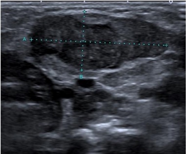
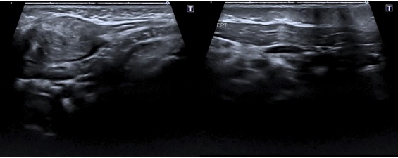
Figure 2: Fusiform thickening of the left sternocleidomastoid muscle. Contralateral sternocleidomastoid muscle with normal thickness and ecostructure 

Fibromatosis colli is the most severe presentation of congenital muscular torticollis and a relatively rare cause of neck swelling in neonates and infants. This pathology occurs in 0.4% of newborns.1 The pathogenesis is still unclear, but it is believed that, after a complicated delivery, there may be neck compression leading to ischemia with consequent damage to muscle fibers.1,2Typically, it appears 2-4 weeks after birth, most commonly following a difficult delivery.3 But even in cases of infants with a normal delivery this diagnosis should be kept in mind. It is usually a self-limiting condition resolving within 4-8 months which requires only physical therapy and conservative management.3,4Fibromatosis colli is classified as a benign fibroblastic and myofibroblastic tumor according to the new WHO classification of Soft Tissue Tumors.5 The diagnosis is possible through the history and physical examination and, normally, it is possible to see the head of the neonate tilted toward the affected side, and there may be an associated jaw rotation to the contralateral shoulder due to sternocleidomastoid muscle contracture. The abnormal position of the neck may lead to facial asymmetry and positional plagiocephaly.6

The differential diagnosis of pediatric neck masses is extensive and should always be considered,7 the most frequent being the differential diagnosis with adenopathy as in our clinical case.

Ultrasound is the diagnosis imaging modality of choice if there is any doubt in the diagnosis. It shows focal or diffuse enlargement of the sternocleidomastoid muscle in a fusiform configuration.8 The mass moves synchronously with the rest of the sternocleidomastoid muscle on real-time sonography. Unlike fibromatosis colli, adenopathy is characterized by ultrasound as a conglomerate tangled mass with normal homogeneously low echogenicity parenchyma and preservation of fatty hyperechoic hilum alongside ovoid morphology.9Maddalozzo and Goldenberg reported that ultrasonography was 100% sensitive for diagnosis when there is a strong suspicion from the clinical history and physical examination of fibromatosis colli.10

The importance of an accurate clinical diagnosis is thus emphasized, and a confident diagnosis implies the need for an ultrasound examination by a radiologist experienced in pediatric imaging with knowledge of the sonographic characteristics of this clinical entity.8

References

1. Nai G, del Hoyo M. Diagnóstico de fibromatosis colli por punção aspirativa por agulha fina: relato de três casos. J Bras Patol e Med Lab. 2005;41:205-7. [ Links ]

2. Alves M, Branco L, Lopes A, et al. Perinatal clinical case. Nascer E Crescer - Birth Growth Med J. 2018;27:263-5. [ Links ]

3. Khalid S, Zaheer S, Wahab S, et al. Fibromatosis colli: A case report. Oman Med J. 2012;27:1-3. [ Links ]

4. Baik G, Blask A, Reilly B. Unilateral neck mass in neonate. J Pediatr. 2018;202:329. [ Links ]

5. Sbaraglia M, Bellan E, Dei Tos AP. The 2020 WHO Classification of soft tissue tumours: news and perspectives. Pathologica. 2021;113:70-84. [ Links ]

6. Wei JL, Schwartz KM, Weaver AL, Orvidas LJ. Pseudotumor of infancy and congenital muscular torticollis: 170 cases. Laryngoscope. 2001;111:688-95. [ Links ]

7. Weinstock MS, Patel NA, Smith LP. Pediatric cervical lymphadenopathy. Pediatr Rev. 2018;39:433-43. [ Links ]

8. Ablin DS, Jain K, Howell L, West DC. Ultrasound and MR imaging of fibromatosis colli (sternomastoid tumor of infancy). Pediatr Radiol. 1998;28:230-3. [ Links ]

9. Bansal AG, Oudsema R, Masseaux JA, Rosenberg HK. US of Pediatric superficial masses of the head and neck. RadioGraphics. 2018;38:1239-63. [ Links ]

10. Maddalozzo J, Goldenberg JD. Pseudotumor of infancy-the role of ultrasonography. Ear Nose Throat J. 1996;75:248-54. [ Links ]

Ethical Disclosures

Financing Support: This work has not received any contribution, grant or scholarship.

Received: July 01, 2022; Accepted: November 14, 2022

Mariana Eiras Dias, Rua das Lameiras nº1, 2565-281 Freiria, Portugal, Email: marianaeirasdias@gmail.com

Conflicts of interest: The authors have no conflicts of interest to declare.

Confidentiality of data: The authors declare that they have followed the protocols of their work center on the publication of data from patients.

Protection of human and animal subjects: The authors declare that the procedures followed were in accordance with the regulations of the relevant clinical research ethics committee and with those of the Code of Ethics of the World Medical Association (Declaration of Helsinki).

© Author(s) (or their employer(s)) and ARP 2023. Re-use permitted under CC BY-NC. No commercial re-use

Creative Commons License This is an open-access article distributed under the terms of the Creative Commons Attribution License