SciELO - Scientific Electronic Library Online

 
vol.35 número1Rabdomiossarcoma de Bexiga em Recém-Nascido com Síndrome de Schimmelpenning índice de autoresíndice de assuntosPesquisa de artigos
Home Pagelista alfabética de periódicos  

Serviços Personalizados

Journal

Artigo

Indicadores

Links relacionados

  • Não possue artigos similaresSimilares em SciELO

Compartilhar


Acta Radiológica Portuguesa

versão impressa ISSN 2183-1351

Acta Radiol Port vol.35 no.1 Lisboa abr. 2023  Epub 30-Abr-2023

https://doi.org/10.25748/arp.28000 

Imagens de Interesse

Pulmonary Cement Embolism

Embolia Pulmonar por Cimento Ósseo

Margarida Morgado1  2 
http://orcid.org/0000-0001-9988-9011

Ana Teresa Vilares1  2 
http://orcid.org/0000-0001-7375-491X

Filipe Barros Alves1  2 
http://orcid.org/0000-0003-1119-9003

Ricardo Castro1 
http://orcid.org/0000-0002-4662-6784

António J. Madureira1  2 
http://orcid.org/0000-0003-2490-1447

1Serviço de Radiologia, Centro Hospitalar Universitário de São João, Porto, Portugal.

2Departamento de Medicina, Faculdade de Medicina da Universidade do Porto, Porto, Portugal


Abstract

Vertebroplasty and kyphoplasty are widely used procedures for patients with symptomatic spinal compression fractures. Cement leakage into the paravertebral veins leading to pulmonary embolism is not an uncommon complication and the risk is increased when treating bone metastases. We present a case and images of a patient with previous vertebral compression fractures from metastatic breast cancer treated with vertebroplasty and kyphoplasty which presented imaging findings consistent with pulmonary cement embolism.

Keywords: Pulmonary embolism; Bone cements; Vertebroplasty; Kyphoplasty; Fractures; Compression.

Resumo

A vertebroplastia e a cifoplastia são procedimentos amplamente utilizados em doentes sintomáticos com fraturas vertebrais compressivas. O extravasamento de cimento ósseo para as veias paravertebrais com consequente embolia pulmonar não é uma complicação incomum e o risco é superior no tratamento de metástases ósseas. Apresentamos um caso clínico com imagens de uma doente com antecedentes de fraturas vertebrais compressivas por metastização óssea de cancro da mama, tratadas com vertebroplastia e cifoplastia, que evidenciou achados imagiológicos compatíveis com embolia pulmonar por cimento ósseo.

Palavras-chave: Embolia pulmonar; Cimentos ósseos; Vertebroplastia; Cifoplastia; Fraturas; Compressão.

Case

A 68-year-old female patient with previous vertebral compression fractures from metastatic breast cancer treated with vertebroplasty and kyphoplasty presented a new incidental thoracic finding on the surveillance computed tomography (CT) scan. Venous phase contrast-enhanced CT revealed high attenuating linear intravascular material within the left pulmonary artery and its posterior, descending and lateral basal branches (Fig. 1). The patient had no respiratory or cardiovascular symptoms.
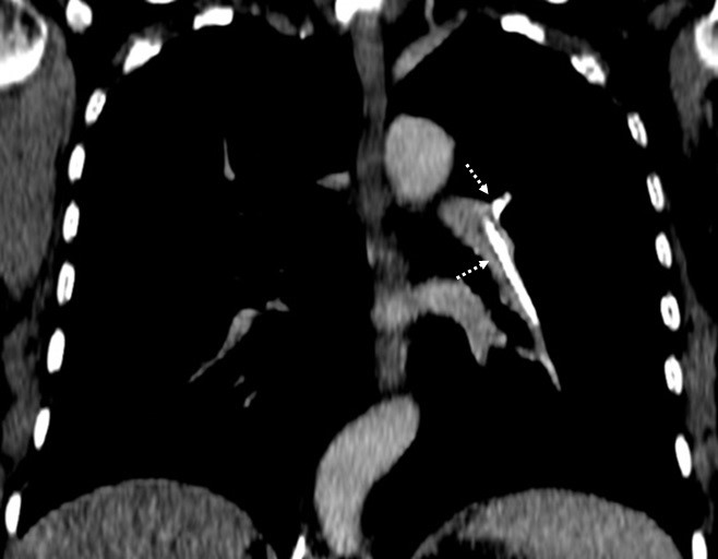
Figure 1: Reconstructed oblique coronal plane venous phase contrast-enhanced CT (mediastinum window) with high attenuating linear intravascular material within the left pulmonary artery and its posterior and descending branches (dashed arrows) consistent with bone cement pulmonary emboli. 

Similar high attenuating linear intravascular material was found within the thoracic anterior external vertebral plexus at the level of T12 and the azygos vein (Fig.2, A); and within the lumbar anterior external vertebral plexus at the level of L5, the left common iliac vein and the inferior vena cava (Fig. 2, B). The vertebral bodies of T11, T12 and L5 were filled with the same high-density material (Fig. 2).
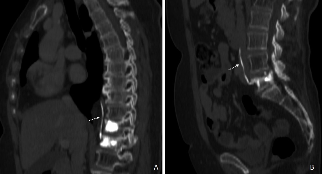
Figure 2: Reconstructed oblique sagittal plane venous phase contrast-enhanced CT (bone window) with high-density material filling the vertebral bodies of T11 and T12 and within the azygos vein (A, dashed arrow) and filling the vertebral body of L5 and within the left common iliac vein and the inferior vena cava (B, dashed arrow) indicating bone cement venous leakage. 
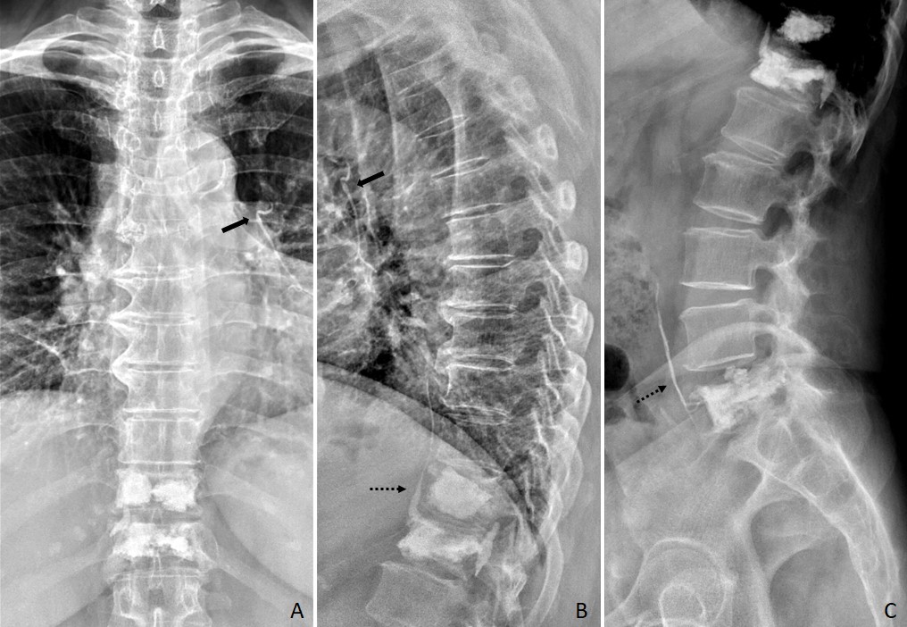
Figure 3: Postoperative thoracic and lumbar spine radiographs after vertebroplasty and kyphoplasty of T11, T12 and L5 demonstrate radio-dense linear opacities on the left peri-hilar region (arrow, A and B) and on the anterior paravertebral region (dashed arrow, B and C) consistent with leaked bone cement within the left pulmonary artery and its posterior and descending branches, the azygos vein, the left common iliac vein and the inferior vena cava. 

These findings were consistent with pulmonary cement embolism after percutaneous vertebroplasty and kyphoplasty.(Fig. 3)

Discussion

Vertebroplasty and kyphoplasty are widely used minimally invasive procedures for patients with symptomatic spinal compression fractures.1,2,3 By injecting polymethyl methacrylate cement into the vertebral body, under image guidance, it provides vertebral stability and immediate pain relief.1,3 Nevertheless, complications such as venous cement leakage into the paravertebral veins leading to pulmonary embolism are not uncommon.1,3 Compression fractures increase venous drainage of the already highly vascularized vertebral bodies3 and the risk of leakage is even higher when treating bone metastases since there is often cortical destruction.1 Furthermore, cement leakage into the inferior vena cava is a significant risk factor for pulmonary embolism.2 Most patients with pulmonary cement embolism are asymptomatic and do not develop any sequelae.1 Nevertheless, when associated with chest pain, dyspnoea, tachycardia or hypoxia, there should be suspicion of pulmonary infarction.2,3

Conclusion

Pulmonary cement embolism is not an unusual complication associated with vertebroplasty and kyphoplasty. Radiologists should be able to recognize the characteristic intravascular and bone findings which allow a definite diagnosis, even when encountered incidentally.

References

1. Choe DH, Marom EM, Ahrar K, Truong MT, Madewell JE. Pulmonary embolism of polymethyl methacrylate during percutaneous vertebroplasty and kyphoplasty. AJR Am J Roentgenol. 2004;183:1097-102. [ Links ]

2. Kim YJ, Lee JW, Park KW, Yeom JS, Jeong HS, Park JM, et al. Pulmonary cement embolism after percutaneous vertebroplasty in osteoporotic vertebral compression fractures: incidence, characteristics, and risk factors. Radiology. 2009;251:250-9. [ Links ]

3. Mansour A, Abdel-Razeq N, Abuali H, Makoseh M, Shaikh-Salem N, Abushalha K, et al. Cement pulmonary embolism as a complication of percutaneous vertebroplasty in cancer patients. Cancer Imaging. 2018;18:5. [ Links ]

Ethical disclosures

Financing Support: This work has not received any contribution, grant or scholarship.

Received: September 04, 2022; Accepted: October 05, 2022

Address Margarida Morgado, Serviço de Radiologia, Centro Hospitalar Universitário de São João, Alameda Prof. Hernâni Monteiro, 4200-319 Porto, Portugal, e-mail: margarida.r.morgado@gmail.com

Conflicts of interest: The authors have no conflicts of interest to declare.

Confidentiality of data: The authors declare that they have followed the protocols of their work center on the publication of data from patients.

Protection of human and animal subjects: The authors declare that the procedures followed were in accordance with the regulations of the relevant clinical research ethics committee and with those of the Code of Ethics of the World Medical Association (Declaration of Helsinki).

© Author(s) (or their employer(s)) and ARP 2023. Re-use permitted under CC BY-NC. No commercial re-use.

Creative Commons License This is an open-access article distributed under the terms of the Creative Commons Attribution License