SciELO - Scientific Electronic Library Online

 
vol.79 issue2Discoid Lupus Erythematosus of the Scalp in a Patient with Systemic Lupus Erythematosus: A Case Report with Complete Hair RegrowthBullous Factitious Disorder Simulating Bullous Pemphigoid: Case Report and Literature Review author indexsubject indexarticles search
Home Pagealphabetic serial listing  

Services on Demand

Journal

Article

Indicators

Related links

  • Have no similar articlesSimilars in SciELO

Share


Revista da Sociedade Portuguesa de Dermatologia e Venereologia

Print version ISSN 2182-2395On-line version ISSN 2182-2409

Rev Soc Port Dermatol Venereol vol.79 no.2 Lisboa June 2021  Epub Oct 01, 2021

https://doi.org/10.29021/spdv.79.2.1305 

Case Report

Isolated Limb Perfusion for the Treatment of Unresectable In-Transit Metastases of Cutaneous Squamous Cell Carcinoma

Perfusão Isolada de Membro no Tratamento de Metástases em Trânsito de Carcinoma Espinocelular

1Dermatology and Venereology Department, Hospital de Santo António dos Capuchos, Centro Hospitalar Universitário de Lisboa Central, Lisboa, Portugal

2Dermatology and Venereology Department, Centro Hospitalar Universitário de Coimbra, Coimbra, Portugal

3General Surgery Department, Instituto Português de Oncologia de Lisboa, Lisbon, Portugal


ABSTRACT

Squamous cell carcinoma (SCC) is the second most common non-melanoma skin cancer and its incidence has been increasing over the past decades. SCC in-transit metastases are rare and predict a poor prognosis.

We present the case of a 69-year-old patient with a right lower leg SCC, surgically excised with free margins. One month later, erythematous to violaceous, firm papules begin to erupt on the right lower leg, evolving to ulcerated nodules over a period of weeks. Homolateral inguinal and iliac nodal metastases were documented by percutaneous biopsy of an inguinal palpable lymph node and later documented by positron emission tomography - computed tomography (PET-CT). Given the unresectability of the disease, regional chemotherapy involving isolated limb perfusion (ILP) with melphalan and tumor necrosis factor-alpha (TNFα) was performed in order to avoid amputation. Macroscopic regression

ensued, with complete resolution of all visible in-transit metastases 12 weeks after perfusion. However, a fatal outcome was observed 4 months later, due to systemic metastases.

ILP is an effective and well-established technique in the treatment of advanced tumors of the extremities. Its main indications are in-transit metastases of malignant melanoma and advanced soft tissue sarcomas. However, studies have shown its effectiveness in other tumors such as locally advanced SCC. Despite the unfortunate outcome, it was possible to avoid amputation of the limb, with complete local disease remission.

KEYWORDS: Amputation; Carcinoma, Squamous Cell; Extremities; Neoplasm Metastasis

RESUMO

O carcinoma espinocelular (CEC) é o segundo cancro cutâneo não-melanoma mais comum e a sua incidência tem aumentado nas últimas décadas. A metastização locoregional é rara e, quando ocorre, associa-se a um pior prognóstico. Apresenta-se o caso de uma doente de 69 anos, com CEC da perna direita, invasivo, moderadamente diferenciado, totalmente excisado cirurgicamente. Após 1 mês, a doente refere o aparecimento de pápulas eritematovioláceas, de consistência dura, que evoluíram para nódulos de 3-4 cm, ulcerados, localizados na perna direita. A invasão ganglionar inguinal e ilíaca homolateral foi documentada através de punção aspirativa de conglomerado adenopático palpável

a esse nível e posteriormente documentado por tomografia de emissão de positrões com TC (PET-TC). Dada a irressecabilidade cirúrgica da doença, foi realizada uma perfusão isolada de membro (PIM) com melfalan e fator de necrose tumoral (TNFα) para evitar a amputação. Foi observada resolução completa de todas as metástases em trânsito 12 semanas após a perfusão. No entanto, 4 meses depois, a progressão sistémica da doença culminou num desfecho fatal.

A PIM é uma técnica eficaz e bem estabelecida no tratamento de tumores avançados das extremidades. As suas principais indicações são as metástases em trânsito de melanoma maligno e sarcomas de partes moles. No entanto, estudos demonstram a sua eficácia no tratamento de outros tumores, como é o caso do CEC localmente avançado. Apesar de não ter alterado o prognóstico da doença, foi possível, com a abordagem instituída, evitar a amputação do membro, atingindo a remissão completa da doença localizada.

PALAVRAS-CHAVE: Amputação; Carcinoma de Células Escamosas; Extremidades; Metástase Neoplásica

INTRODUCTION

Non-melanoma skin cancer (NMSC) represents the most common type of cancer in caucasians, with an incidence that has been steadily increasing worldwide. Basal cell carcinoma and squamous cell carcinoma (SCC) account for the majority of NMSC cases.1,2The most commonly affected anatomic regions are sun-exposed areas like the head and neck, although a large proportion are located on the limbs.3 Extension of the disease will determine the most suitable treatment approach. Multiple treatment strategies are available for localized disease, from wide surgical excision - usually the most common treatment - to Mohs micrographic surgery, cryosurgery or radiotherapy. However, when facing a locally advanced and/ or widespread disease, with clinical and histopathological features associated with bad outcomes, surgical excision with adequate free margins may not be appropriate, leading to serious loss of limb function or even limb amputation.4

Isolated limb perfusion (ILP) is a well-established technique used for the treatment of unresectable tumors of the extremities.2,4It was first introduced in clinical practice by Creech and Krementz, in 1957, in New Orleans, United States of America, in a patient with in-transit metastases of malignant melanoma, who achieved a complete response.5 Nowadays, ILP is mainly used for the treatment of in-transit metastases of malignant melanoma and advanced soft tissue sarcomas of the extremities.4,6However, recent studies have shown ILP’s efficacy in rarer tumours confined to the extremities - locally advanced SCC, cutaneous lymphomas, Merkel cell carcinomas and desmoid tumors.7 ILP has proven to be a safe and effective limb-saving strategy without the systemic toxicity of conventional chemotherapy.4

CASE REPORT

We present the case of a 69-year-old female patient with a history of kidney transplant 10 years before, on immunosuppressive therapy with prednisolone, mycophenolate mofetil and everolimus.

The patient was admitted to the Dermatology Department with a 6-month history of a growing ulcerated nodule on her lower right leg, measuring 5 cm. Wide surgical excision was performed followed by closure with a full-thickness skin graft. Histopathology revealed a completely excised, invasive, moderately differentiated SCC, without perineural or lymphovascular invasion. Four weeks later, multiple erythematous-violaceous papules begin to erupt on the lower right leg in a linear pattern. Enlarged homolateral inguinal lymph nodes were also noticed on clinical examination. Skin and lymph node biopsies were performed and were positive for cutaneous and lymph node metastases of SCC, respectively. Staging PET-CT showed hypermetabolic activity in these locations. On re-evaluation, 4 weeks later, the lesions had evolved to 3-4 cm, exophytic, ulcerated, haemorrhagic nodules, located on the anterior and posterior surface of the lower leg (Fig. 1). Given the regional spread of the disease with locoregional metastases, the patient was treated with ILP, consisting of a combination of melphalan and TNFα. An iliac perfusion was performed with temperatures ranging from 39.5ºC to 40ºC, using 90 mg of melphalan (10 mg/L limb volume) and 1 mg of TNFα. Inguinal and ilio-obturator lymph node dissection was performed at the same time, without immediate intercurrences. A total of 11 lymph nodes were excised, 10 of which had metastases from SCC.

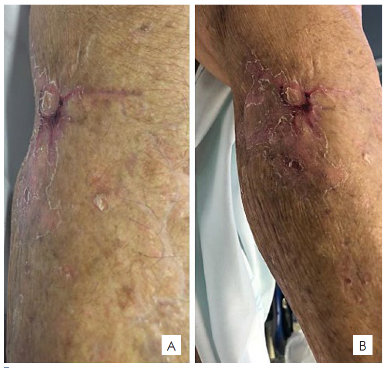
As soon as 1 week after the procedure, partial disease regression was noticed, with a reduction in lesions size and erythema (Fig. 2A) with more significant reduction recorded at week 4 (Fig. 2B). Complete resolution of all in-transit metastases was observed at week 12, leaving only a residual scar on the anterior surface of the leg (Fig. 3). The patient did not experience any local or systemic toxicity. Nevertheless, 4 months later, systemic progression of the disease was reported on the PET-TC, showing hypermetabolic activity at multiple locations. Unfortunately, the patient died 4 months later.

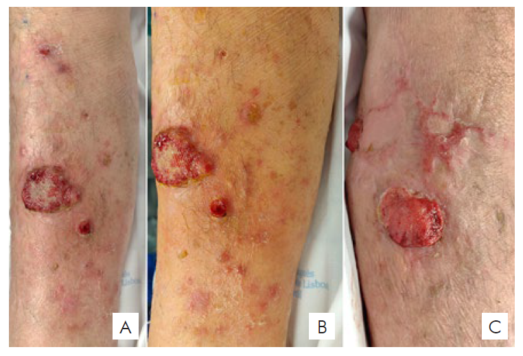
Figura 1 Multiple erythematous and violaceous papulonodules covered with haemorrhagic crusts, located on the anterior (A, B), lateral and posterior (C) aspect of the right lower leg. 

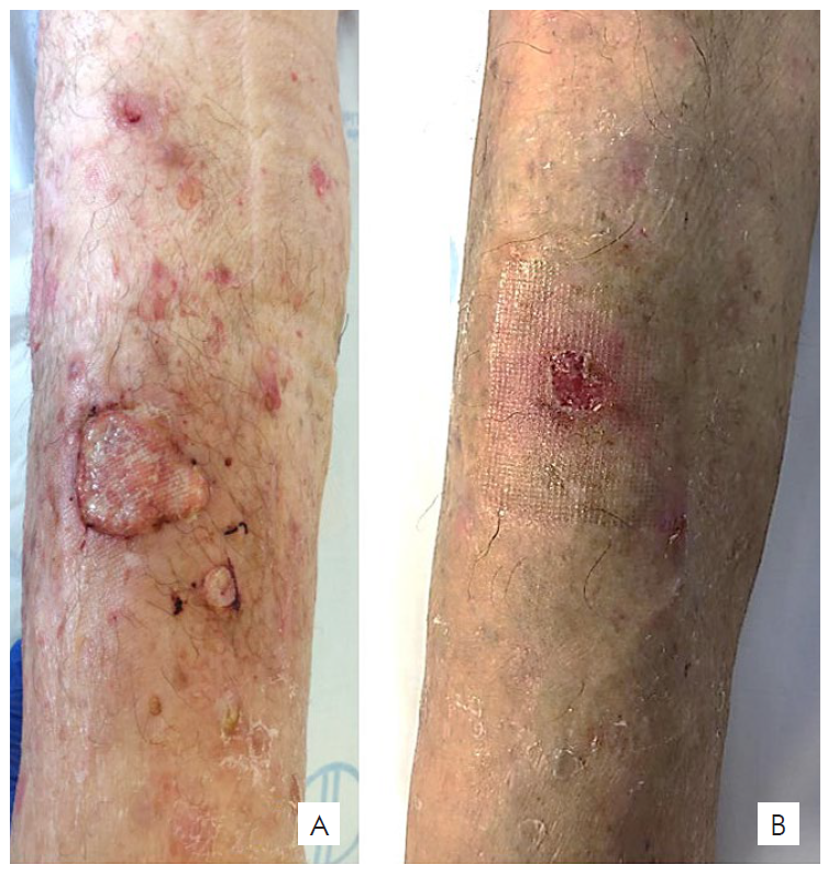
Figura 2 One week post-procedure - reduction in size and erythema of the cutaneous lesions (A); Four weeks post-procedure - 1x1 cm erosion on the anterior aspect of the right lower leg with complete resolution of the remaining lesions (B). 

Figura 3 Twelve weeks post-procedure. Complete resolution of the cutaneous lesions with residual atrophic scaring. 

DISCUSSION

SCC accounts for most non-melanoma skin cancer-related metastatic disease and deaths.8 Once an invasive/aggressive SCC is diagnosed, it must be staged as the risk for metastases is reported to be approximately 49 and 2 to 3 times higher in immunosuppressed individuals.10 Cutaneous in-transit and regional lymph node metastases are the most common metastatic presentation.8 Treatment modalities described for advanced SCC at any site include conventional cytotoxic chemotherapy, newer systemic therapies, radiotherapy and combinations of these modalities.4 The ILP technique has been extensively described. Its purpose is to allow local administration of high-dose chemotherapy while minimizing serious systemic side effects.3,11

Briefly, after heparinization, the surgeon identifies and cannulates the main vessels of the limb, in this case the right iliac vessels, which were clamped proximally. With the use of an extracorporeal circulation pump, chemotherapy with melphalan and TNFα is infused into the limb. The technique is based on the synergistic antineoplastic effect of chemotherapeutic drugs and high temperature,7 which should be maintained between 38°C and 40°C, measured with two thermometers placed in subcutaneous tissue and muscle. Systemic and regional toxicity should be assessed during perfusion with radiomarked albumin infused on the systemic circulation and a gamma probe.2,7,11,12

Several reports show excellent response rates with TNFα and melphalan- ILP for both malignant melanoma in-transit metastases or locally advanced soft tissue sarcomas.3,6,7However, there is not much evidence on its efficacy when it comes to non-melanoma skin cancer, such as SCC. Veld et al recently reported a case series of 30 patients with locally advanced SSC treated with ILP. The overall response rate was 81%, with 16 patients having a complete response (59%). Limb salvage rate was 80% and the overall 2-year survival was 67%. The authors suggest that ILP should be considered as an option in patients with locally advanced SCC.4 Belgrano et al report two patients with SCC submitted to ILP, with disease stabilization in one patient and a complete response in the other. However, both experienced local progression of the disease within 4-7 months.7

In this case of a SCC with locoregional metastases, the regression of all in-transit metastases was considered an indicator of a successful outcome, mitigating the need for limb amputation. However, despite its regional efficacy, ILP is unlikely to have a great impact on survival, as shown in several large prospective studies on melanoma and locally advanced soft tissue tumors.6,13,14The widespread disease observed later in this patient might have been a result of systemic metastases that were undetected by imaging and clinical evaluation.

In conclusion, in this patient, ILP was an effective and well tolerated limb-saving palliative treatment. However, it did not have any impact on the overall prognosis. This technique can be regarded as a therapeutic option in the management of multiple, locally advanced, drug-resistant tumors of the limbs, when surgical therapy is not feasible.

Presentations/ Apresentações

This case has been presented as a poster on “XIX Congresso Nacional de Dermatologia e Venereologia”, Porto, 6-8th December of 2019.

REFERENCES

1. Samarasinghe V, Madan V. Nonmelanoma Skin Cancer. J Cutan Aesthet Surg. 2012;5:3-10. [ Links ]

2. Jiménez Herevia AE, Tavares de la Paz LA, Hinojosa Ugarte D, Nieves Condoy J. Isolated limb perfusion on nonmelanoma skin cancer for limb salvage: a series of four cases. Cureus. 2020;12:e9998. doi: 10.7759/cureus.9998. [ Links ]

3. Alegre JF, Duarte M, Sureda González M, Bretcha Boix P, Dussan C, Ballester A, et al. Tratamiento regional del sarcoma y melanoma localmente avanzado de la extremidad mediante la perfusión aislada con tumor necrosis factor a y melfalan asociada a hipertermia. Cir Esp. 2012;90:114-20. [ Links ]

4. Huis In 't Veld EA, Grünhagen DJ, Deroose JP, Nijsten TC, Wouters MW, Verhoef C, et al. Isolated limb perfusion for unresectable extremity cutaneous squamous cell carcinoma; an effective limb saving strategy. Br J Cancer. 2018;119:429-34. doi: 10.1038/s41416-018-0149-z. [ Links ]

5. Creech O, Krementz ET, Ryan RF, Winblad JN. Chemotherapy of cancer: regional perfusion utilizing an extracorporeal circuit. Ann Surg. 1958;148:616-32. [ Links ]

6. Bhangu A, Broom L, Nepogodiev D, Gourevitch D, Desai A. Outcomes of isolated limb perfusion in the treatment of extremity soft tissue sarcoma: a systematic review. Eur J Surg Oncol. 2013;39:311-9. doi: 10.1016/j.ejso.2012.12.018. [ Links ]

7. Belgrano V, Ben-Shabat I, Bergh P, Bagge RO. Isolated limb perfusion as a treatment option for rare types of tumours. Int J Hyperthermia. 2016;32:595-9. doi: 10.1080/02656736.2016.1188999. [ Links ]

8. Combalia A, Carrera C. Squamous cell carcinoma: an update on diagnosis and treatment. Dermatol Pract Concept. 2020;10:e2020066. [ Links ]

9. Brantsch KD, Meisner C, Schönfisch B, Trilling B, Wehner-Caroli J, Röcken M, et al. Analysis of risk factors determining prognosis of cutaneous squamous-cell carcinoma: a prospective study. Lancet Oncol. 2008;9:713-20. [ Links ]

10. Cooper JZ, Brown MD. Special concern about squamous cell carcinoma of the scalp in organ transplant recipients. Arch Dermatol. 2006;142:755-8. [ Links ]

11. Ponte P, Moniz JV, Farricha V, Weinholtz JB. Merkel cell carcinoma: an unusual indication for isolated limb perfusion. Dermatol Online J. 2008;14:6. [ Links ]

12. Olieman AF, Liénard D, Eggermont AM, Kroon BB, Lejeune FJ, Hoekstra HJ, et al. Hyperthermic isolated limb perfusion with tumor necrosis factor alpha, interferon gamma, and melphalan for locally advanced nonmelanoma skin tumors of the extremities: a multicenter study. Arch Surg. 1999;134:303-7. [ Links ]

13. Aloia TA, Grubbs E, Onaitis M, Mosca PJ, Cheng TY, Seigler H, et al. Predictors of outcome after hyperthermic isolated limb perfusion: role of tumor response. Arch Surg. 2005;140):1115-20. [ Links ]

14. Koops HS, Vaglini M, Suciu S, Kroon BB, Thompson JF, Göhl J, et al. Prophylactic isolated limb perfusion for localized, high-risk limb melanoma: results of a multicenter randomized phase III trial. European Organization for Research and Treatment of Cancer Malignant Melanoma Cooperative Group Protocol 18832, the World Health Organization Melanoma Program Trial 15, and the North American Perfusion Group Southwest Oncology Group-8593. J Clin Oncol. 1998;16:2906-12. [ Links ]

1© Author(s) (or their employer(s)) 2021 SPDV Journal. Re-use permitted under CC BY-NC. No commercial re-use. © Autor (es) (ou seu (s) empregador (es)) 2021 Revista SPDV. Reutilização permitida de acordo com CC BY-NC. Nenhuma reutilização comercial

Received: November 18, 2020; Accepted: January 05, 2021

Corresponding Author: Joana Barbosa Address: Serviço de Dermatologia do Hospital de Santo António dos Capuchos, Alameda Santo António dos Capuchos, 1169-050 Lisboa, Portugal E-mail:joana.barbosa@chlc.min-saude.pt

Conflicts of Interest: The authors have no conflicts of interest to declare. Financing Support: This work has not received any contribution, grant or scholarship. Confidentiality of Data: The authors declare that they have followed the protocols of their work center on the publication of data from patients. Patient Consent: Consent for publication was obtained. Provenance and Peer Review: Not commissioned; externally peer reviewed. Conflitos de Interesse: Os autores declaram a inexistência de conflitos de interesse na realização do presente trabalho. Suporte Financeiro: Não existiram fontes externas de financiamento para a realização deste artigo. Confidencialidade dos Dados: Os autores declaram ter seguido os protocolos da sua instituição acerca da publicação dos dados de doentes. Consentimento: Consentimento do doente para publicação obtido. Proveniência e Revisão por Pares: Não comissionado; revisão externa por pares

Creative Commons License This is an open-access article distributed under the terms of the Creative Commons Attribution License