SciELO - Scientific Electronic Library Online

 
vol.79 número1Utilidade da Ecografia Cutânea na Celulite Dissecante: A Propósito de 2 Casos ClínicosUretrite Masculina Causada por Trichomonas vaginalis índice de autoresíndice de assuntosPesquisa de artigos
Home Pagelista alfabética de periódicos  

Serviços Personalizados

Journal

Artigo

Indicadores

Links relacionados

  • Não possue artigos similaresSimilares em SciELO

Compartilhar


Revista da Sociedade Portuguesa de Dermatologia e Venereologia

versão impressa ISSN 2182-2395versão On-line ISSN 2182-2409

Rev Soc Port Dermatol Venereol vol.79 no.1 Lisboa mar. 2021  Epub 15-Jun-2021

https://doi.org/10.29021/spdv.79.1.1269 

Case Reports

Pigmented Epithelioid Melanocytoma: Case Report

Melanocitoma Epitelioide Pigmentado: Relato de Caso

Eduardo Scardazzi Silva Ragni1 

Marcel Arakaki Asato1  2 

Estela Mari Sandini1 

Lucas Basmage Pinheiro Machado1 

Sylka Rebelato Toppan1 

1 Universidade Federal do Mato Grosso do Sul - Faculdade de Medicina - Departamento de Dermatologia, Campo Grande, , Brasil

2Universidade Federal do Mato Grosso do Sul - Faculdade de Medicina - Departamento de Patologia, Campo Grande, Brasil


Abstract

Abstract: Pigmented epithelioid melanocytoma is a rare, newly described melanocytic tumor that encompasses lesions previously classified as animal type melanomas and epithelioid blue nevus of the Carney complex. Pigmented epithelioid melanocytoma is a specific clinicopathological entity with particular clinical presentation and histological features. We present the case of a 5 year old female patient with a heavily pigmented papule on her right thigh that showed histological findings compatible with pigmented epithelioid melanocytoma and discuss the relevance /clinical significance of sentinel lymph node biopsy as a staging procedure in this particular neoplasm.

Keywords: Child; Melanoma; Nevus, Blue; Nevus, Pigmented; Skin Neoplasms

Resumo:

Melanocitoma epitelioide pigmentado é um tumor raro, recentemente descrito que engloba lesões previamente classificadas como melanomas “tipo animal” e nevo azul epitelioide do complexo de Carney. Melanocitoma epitelioide pigmentado é uma entidade clinicohistopatológica específica com apresentação clínica e características histopatológicas particulares. Apresentaremos o caso de uma paciente do sexo feminino de 5 anos de idade com uma pápula densamente pigmentada em sua coxa direita que mostrou achados histológicos compatíveis com Melanocitoma epitelioide pigmentado e discutiremos a relevância/ significância clínica da biópsia de linfonodo sentinela como parte do processo de estadiamento dessa neoplasia em particular.

Palavras-chave: Criança; Melanoma; Neoplasias da Pele; Nevo Azul; Nevo Pigmentado

Introduction

Pigmented epithelioid melanocytoma (PEM) is a rare melanocytic tumor composed of pigmented epithelioid and dendritic melanocytes with large nuclei.1This histologic entity encompasses tumors previously described as epithelioid blue nevus of the Carney complex2,3and "animal-type melanomas". 2,4

Clinically, it presents as a slowly growing, densely pigmented papule or nodule on extremities, trunk and head.1This tumor is more common in young adults and children but can occur at any age.2

Most cases are sporadic1 and a small subset of cases is associated with the Carney complex.2 Carney’s complex is a familial syndrome characterized by blue naevi, lentigines, myxomas and rare tumors such as psammomatous melanotic schwannoma.3

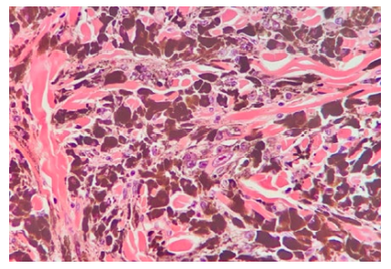
Histological findings of PEM include: wedge-shaped symmetrical configuration; central increased cellularity; epithelioid cells, dendritic cells and melanophages.1 Clusters of large multinucleated cells with vesicular nuclei and macronucleoli (Reed-Sternberg-like) are the most distinct finding in PEM.1

The treatment comprises of simple excision. Although Ludgate et al and Zembowics et al reported a positive sentinel lymph node biopsy (SLNB), respectively in 47%5 and 46%,2 there is no evidence to recommend that sentinel lymph node sampling in primary cutaneous PEM has therapeutic or prognostic value.1

This case report describes the diagnosis of PEM in a 5-year-old patient presenting with classic clinical features and compatible histological findings.

Case Report

The patient is a female 5-year-old from Campo Grande in the state of Mato Grosso do Sul, Brazil. At a Dermatology consultation her mother expressed concern over a dark papule that had appeared on her daughter's right thigh 2 years prior and was slowly growing in size. The lesion was asymptomatic. No palpable lymph node was noted. There was no history of frequent sun exposure.

The patient had no prior medical conditions, was not using any medication and had never undergone any skin biopsy procedures. There was no personal or family history of skin cancer.

A 7 mm hyperpigmented bluish papule was observed on the posterior right thigh. On dermoscopy the lesion showed a continuous blue-gray blotch of pigment with no other visible structures.

The lesion was promptly excised. Histopathology showed a circumscribed, symmetrical, wedge-shaped dermal proliferation composed predominantly of pigmented epithelioid cells and dendritic cells. The cells had large nuclei and prominent nucleoli. In some areas multinucleated cells were found. No mitotic figures were identified.

The clinical and histological findings were compatible with PEM.

Figure 1: Dermoscopy image of the lesion showing a continuous blue-gray blotch of pigment 

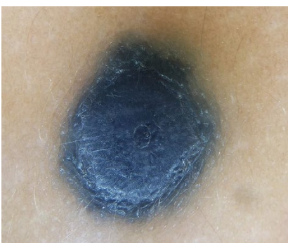
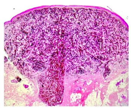
Figure 2: Symmetrical, wedge-shaped dermal proliferation composed predominantly of pigmented epithelioid and dendritic cells (H&E x100). 

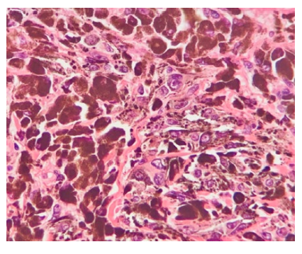
Figure 3: Pigmented epithelioid and dendritic cells with large nuclei and prominent nucleoli (H&E x400). 

Figure 4: In some areas multinucleated cells were found (H&E x400). 

Discussion

Sentinel lymph node (SLN) involvement observed in the majority of PEMs6 along with the tumor once being categorized as an "animal-type" melanoma2 prompted most previous reports to include SLN sampling as a diagnostic procedure. However, the frequent SLN involvement in PEMs does not have the same adverse prognostic significance as in conventional melanoma2,6-8and the positive rate in cases undergoing SNLB may be over-estimated due to a more progressive clinical course or deeper tumor cell invasion.9

Another theory is that PEMs may be part of the neoplasms known as "benign metastasizing” tumors.8 These particular kinds of tumors include both cutaneous and non-cutaneous lesions such as benign melanocytic naevi, endometriosis and metastasizing leiomyomas.8

Additionally, based on current studies, a positive sentinel lymph node sampling appears to present little important evidence of the lesion's biological potential and its risk of developing clinically significant metastatic disease.10Taking that into consideration it can be argued that SLN sampling should not be performed in every patient with PEM.8

There are no clearly established criteria for performing SLNB in PEM patients9 and no histological criteria predictive of potential metastatic behavior of the tumor.2,10Current studies suggest that PEM is an indolent melanocytic tumor and long-term follow-ups have not revealed disease-specific lethal cases regardless of tumor deposits in sentinel lymph node sampling.1

Sentinel lymph node sampling was not performed in this case due to lack of evidence proving diagnostic or prognostic value of this practice on this kind of tumor.1A complementary wide surgical excision was performed and the patient will go through a long follow-up period.

In conclusion, while it is considered to be an indolent tumor, PEM patients should undergo a long follow-up period and further studies are needed on whether SLNB should be performed as a staging procedure in every case of PEM.

References

1. Zembowicz A, Cohen JN, LeBoit PE. Pigmented Epithelioid Melanocytoma. In: Busam KJ, Gerami P, Scolyer RA, editor. Pathology of Melanocytic Tumors. Amsterdam: Elsevier; 2019. p.124-9. [ Links ]

2. Zembowicz A, Carney JA, Mihm CM Jr. Pigmented epithelioid melanocytoma, a low grade melanoma indistinguishable from animal type melanoma and epithelioid blue nevus. Am J Surg Pathol. 2004;28:31-40. doi: 10.1097/00000478-200401000-00002. [ Links ]

3. Carney JA, Ferreiro JA. The epithelioid blue nevus. A multicentric familial tumor with important associations, including cardiac myxoma and psammomatous melanotic schwannoma. Am J Surg Pathol. 1996;20:259-72. [ Links ]

4. Requena LM. Animal type melanoma: a report of a case with balloon-cell change and sentinel lymph node metastasis.. Am J Dermatopathol. 2001;23:341-6. [ Links ]

5. Ludgate MW, Fullen DR, Lee J, Rees R, Sabel MS, Wong SL, Johnson TM. Animal-type melanoma: a clinical and histopathological study of 22 cases from a single institution. Br J Dermatol 2010; 162: 129-36. doi: 10.1111/j.1365-2133.2009.09271.x. [ Links ]

6. Mandal RV, Murali R, Lundquist KF, Ragsdale BD, Heenan P, McCarthy SW, et al. Pigmented epithelioid melanocytoma: favorable outcome after 5-year follow-up. Am J Surg Pathol. 2009;33:1778-82. doi: 10.1097/PAS.0b013e3181b94f3c. [ Links ]

7. Zembowicz A, Knoepp SM, Bei T, Stergiopoulos S, Eng C, Mihm MC, et al. Loss of expression of protein kinase a regulatory subunit 1alpha in pigmented epithelioid melanocytoma but not in melanoma or other melanocytic lesions. Am J Surg Pathol. 2007;31:1764-75. oi: 10.1097/PAS.0b013e318057faa7. [ Links ]

8. Lim C, Murali R, McCarthy SW, Krivanek J, Scolyer RA. Pigmented epithelioid melanocytoma: a recently described melanocytic tumour of low malignant potential. Pathology. 2010;42:284-6. doi: 10.3109/00313021003631213. [ Links ]

9. Cheng PS, Chuang SS, Kuo TT, Lai FJ. Pigmented epithelioid melanocytoma: Report of a case and review of 173 cases in the literature. Dermatol Sinica. 2012;30:57-61. doi: 10.1016/j.dsi.2011.09.011. [ Links ]

10. Gavriilidis P, Michalopoulou I, Chatzikakidou K, Nikolaidou A. Pigmented epithelioid melanocytoma: a new concept encompassing animal-type melanoma and epithelioid blue nevus. BMJ Case Rep. 2013;2013:bcr-2013-008865. doi: 10.1136/bcr-2013-008865. [ Links ]

Responsabilidades ÉticasConflitos de Interesse: Os autores declaram a inexistência de conflitos de interesse na realização do presente trabalho. Suporte Financeiro: Não existiram fontes externas de financiamento para a realização deste artigo. Confidencialidade dos Dados: Os autores declaram ter seguido os protocolos da sua instituição acerca da publicação dos dados de doentes. Consentimento: Consentimento do doente para publicação obtido. Proveniência e Revisão por Pares: Não comissionado; revisão externa por pares

Ethical Disclosures Conflicts of Interest: The authors have no conflicts of interest to declare. Financing Support: This work has not received any contribution, grant or scholarship. Confidentiality of Data: The authors declare that they have followed the protocols of their work center on the publication of data from patients. Patient Consent: Consent for publication was obtained. Provenance and Peer Review: Not commissioned; externally peer reviewed

3© Author(s) (or their employer(s)) 2021 SPDV Journal. Re-use permitted under CC BY-NC. No commercial re-use. © Autor (es) (ou seu (s) empregador (es)) 2021 Revista SPDV. Reutilização permitida de acordo com CC BY-NC. Nenhuma reutilização comercial

Received: September 03, 2020; Accepted: November 01, 2020

Corresponding Author: Eduardo Scardazzi Silva Ragni Adress: Ambulatório de Dermatologia da Universidade Federal de Mato Grosso do Sul - Brasil; Avenida Senador Filinto Müler, 355, Vila Ipiranga, Campo Grande, MS, 79080-190, Brasil E-mail: eduardo_rss@hotmail.com

Creative Commons License This is an open-access article distributed under the terms of the Creative Commons Attribution License