SciELO - Scientific Electronic Library Online

 
vol.78 número3Eritema Periocular Induzido pelo Álcool: Efeito Adverso Raro de TacrolimusMalformações Venosas e Arteriovenosas do Lábio: Um Diagnóstico Diferencial Problemático índice de autoresíndice de assuntosPesquisa de artigos
Home Pagelista alfabética de periódicos  

Serviços Personalizados

Journal

Artigo

Indicadores

Links relacionados

  • Não possue artigos similaresSimilares em SciELO

Compartilhar


Revista da Sociedade Portuguesa de Dermatologia e Venereologia

versão impressa ISSN 2182-2395versão On-line ISSN 2182-2409

Rev Soc Port Dermatol Venereol vol.78 no.3 Lisboa set. 2020  Epub 01-Set-2021

https://doi.org/10.29021/spdv.78.3.1248 

Caso Clínico

Case of Flexural Exanthema as a Presenting Sign for COVID-19

Um Caso de Exantema Flexural como Sinal de Apresentação de COVID-19A

Raquel Oliveira1 

Margarida Gonçalo2  3 
http://orcid.org/0000-0001-6842-1360

Carlos Faria4 

David Donaire1 

Benilde Barbosa1 

José Carlos Cardoso2  3 

Maria José Julião4 

José Moura1  5 

Armando Carvalho1  5 

1Department of Internal Medicine, Centro Hospitalar e Universitário de Coimbra, Coimbra, Portugal

2Department of Dermatology, Centro Hospitalar e Universitário de Coimbra, Coimbra, Portugal

3Clinic of Dermatology, Faculty of Medicine, University of Coimbra, Coimbra, Portugal

4Department of Pathology, Centro Hospitalar e Universitário de Coimbra, Coimbra, Portugal

5University Clinic of Internal Medicine, Faculty of Medicine, University of Coimbra, Coimbra, Portugal


ABSTRACT

We report a case of a 84-year-old male hospitalized for bacterial pneumonia who, during hospitalization, developed a flexural exanthema in parallel with a positive swab for SARS-CoV-2. Supportive therapy was instituted, the rash disappeared in 7 days and the patient fully recovered. At the same time, two other cases of SARS-CoV-2 infection occurred in the same ward.

Histopathology and immunohistochemistry of a skin biopsy showed a scarce predominantly perivascular lymphocytic infiltration in the upper dermis, predominantly by CD4+ T cells, a slight epidermotropism, spongiosis and focal parakeratosis, compatible with a viral exanthema or a maculopapular drug eruption. Patch testing with possible culprit drugs were negative.

We seek to add value in understanding all the manifestations of SARS-CoV-2 infection and to draw attention to the importance of early identification of skin manifestations in association with COVID-19.

KEYWORDS: Coronavirus Infections; COVID-19; Exanthema; SARS-CoV-2.

RESUMO

Os autores descrevem o caso de um homem de 84 anos hospitalizado por pneumonia bacteriana que foi posteriormente diagnosticado com COVID-19 e observado com exantema flexural. Com terapêutica de suporte o rash desapareceu em 7 dias e o doente recuperou completamente. Na mesma altura surgiram dois outros casos de infeção por SARS-CoV-2 na mesma enfermaria.

O estudo histopatológico e imunopatológico de biópsia de pele mostrou um infiltrado inflamatório perivascular de predomínio linfoplasmocitário ligeiro na derme superficial, predominantemente CD4+, um ligeiro epidermotropismo, espongiose e paraqueratose focal, compatível com um exantema viral ou uma erupção maculopapular medicamentosa.

Os autores pretendem contribuir para uma melhor compreensão das manifestações da infeção pelo SARS-CoV-2 e alertar para a importância da identificação precoce de alterações cutâneas possivelmente associadas a COVID-19.

PALAVRAS-CHAVE: COVID-19; Exantema; Infecções por Coronavírus; SARS-CoV-2

INTRODUCTION

The COVID-19 pandemic has had a major worldwide impact, even though we still know little about this disease.

Along with the typical respiratory symptoms of viral infections, various cutaneous manifestations have also been associated with SARS-CoV-2, so Dermatology has made na enormous contribution to this pandemic.1

Two main studies described more than 50% of cutaneous manifestations appearing after hospitalization,2,3

Lesions were mostly a skin rash (>70%) affecting mainly the trunk and upper limbs, which was only mildly pruritic and self-limited (2-5 days duration). But a wide range of skin manifestations has been associated with COVID-19: petechial, erythematous, urticarial, vesicular eruptions, transient livedo reticularis, Raynaud phenomena, annular, erythema-multiforme and chilblain-like lesions.4-9

Getting to know SARS-CoV-2 manifestations and its pathogenesis is one of the cornerstones of investigation since it directs the study of possible treatments. Furthermore, it is of utmost importance to raise awareness of COVID-19 skin manifestations as primary manifestations.

CASE REPORT

A 84-year-old male was taken to the emergency department (ED) as he was found lying on the floor of his home with associated sphincter incontinence. His clinical history was notable for dyslipidemia, hypertension and an ischemic stroke. Since 2016, he had thrombocytopenia of unknown cause. He was under daily treatment with amlodipine 5 mg, furosemide 80 mg, and simvastatin 20 mg. No drug allergies were reported.

At the ED, he had a 7-point Glasgow Coma Scale, was hypothermic (tympanic temperature 29.9ºC) and had purulent respiratory secretions which needed to be aspirated.

There were no acute findings on brain computed tomography (CT) scan or electroencephalogram. He had leucocytosis (14.2 x 10⁹/L) with neutrophilia (12.72 x 10⁹/L), lymphocytopenia (0.47 x 10⁹/L), thrombocytopenia (104 x 109/L), high C-reactive protein (4.43 mg/dL) and D-dimers (671 ng/dL). A chest radiography showed bilateral hilar reinforcement and areas of hypotransparency already present in previous exams. Chest CT showed bilateral thickening of the bronchial walls, an area of parenchymal densification in the apical segment of the right upper lobe, and fibro-atelectatic areas in the lower lobes. A nasopharyngeal swab for SARS-CoV-2 was negative. He was then admitted to a medical ward with the diagnosis of community-acquired pneumonia and hypoxemic respiratory insufficiency.

The patient completed 5 days of azithromycin 500 mg IV and 8 days of ceftriaxone 1000 mg IV with recovery of consciousness, resolution of respiratory failure and normalization of imaging and analytical parameters, except for thrombocytopenia.

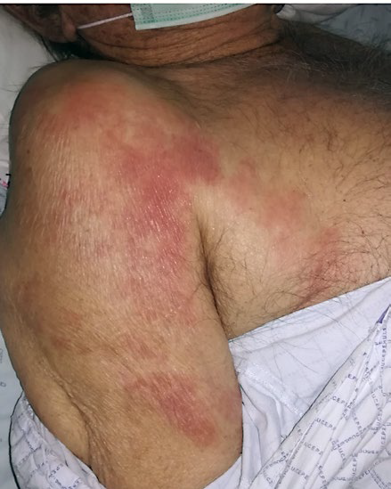
Nevertheless, on day 7 of hospitalization, with no associated respiratory distress or other relevant systemic symptoms, he developed a non-pruritic symmetric maculopapular rash on the trunk and arms, next to axillary folds, with red confluent erythematous lesions that extended to the shoulder and flexures. On the following day, cutaneous lesions progressed to the cervical region and upper trunk (Fig. 1), subsequently stabilizing until resolution within 7 days of onset. Lower limbs, face, palmoplantar regions and mucous membranes were spared. Concomitantly, a second SARS-CoV-2 nasopharyngeal swab was positive and there was a slight increase of inflammatory serum parameters, namely leukocytes (10.4 x 10⁹/L), neutrophils (7.95 x 10⁹/L) and C-reactive protein (7.47 mg/dL), but prothrombin time, activated partial thromboplastin, renal, hepatic and thyroid function tests, antinuclear (ANA) and anti-neutrophil cytoplasmic antibodies (ANCA) were normal or negative. Two other patients in the same ward, with no exposure to antibiotics, had a similar rash and has a positive nasopharyngeal swab for SARS-CoV-2.

Figure 1 Maculopapular rash in the right axillary region. 

For the differential diagnosis of this patient a non-immediate drug eruption related to the antibiotics and a viral exanthema related with SARS-CoV-2 infection were considered.

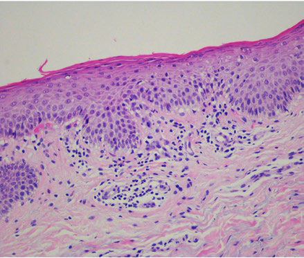
A punch biopsy performed on the patient’s right arm showed mild parakeratosis, focal spongiosis and lymphocyte exocytosis. Vasodilation was seen in the upper dermis with a predominantly perivascular lymphocytic infiltrate, no eosinophils and very rare extravasated red blood cells.

There were no signs of vasculitis (Fig. 2). Immunohistochemistry revealed almost exclusively T cells, predominantly CD4+, with only mild expression of CD8+.

The rash resolved in a week with no specific treatment and the patient recovered fully from COVID-19 only with supportive therapy. Six weeks after recovery, a patch test with several systemic antibiotics, including azithromycin and ceftriaxone at 10% pet (Chemotechnique Diagnostics, Vellinge Sweden), performed according to the ESCD recommendations, 10 was negative on day 2 and day 4.

Figure 2 Perivascular lymphocytic infiltrate in the upper dermis with vasodilatation but no signs of haemorrhage or vasculitis and focal areas of spongiosis and exocytosis in the epidermis. (H&E 200x) . 

DISCUSSION

A symmetric maculopapular rash of the neck, trunk and upper limbs, occasionally also with a flexural distribution, that appears concomitantly with the identification of SARS-CoV-2 in the nasopharyngeal mucosae has been described by other authors11 and may be the initial manifestation of the viral infection. However, in this case, we cannot absolutely exclude the pathogenic role of the antibiotics taken for bacterial pneumonia, even though negative patch tests and the scarcity of eosinophils in the skin biopsy make it unlikely. Otherwise, as reported for Epstein-Barr virus and cytomegalovirus, the simultaneous viral infection and antibiotic intake may have enhanced the cutaneous rash.

Pathophysiology of these cutaneous lesions in COVID-19 is still unknown. A skin biopsy showing vasodilation and a perivascular infiltrate of CD4+ T cells in the upper dermis with a slight epidermotropism has been shown in other viral exanthema and is similar to the ones found in other skin biopsies from COVID-19 patients.6,12

SARS-CoV-2 has been widely suspected as the aetiology of different cutaneous manifestations in infected patients, with maculopapular and urticarial exanthema mostly associated with an indolent course with minimal or even absent fever or respiratory symptoms. However, patients and health professionals are not fully aware that exanthema may be an initial manifestation of the infection, and that these patients are likely contagious.

In summary, maculopapular exanthema has been described in patients who tested positive for SARS-CoV-2, but more studies are needed to clear the pathomechanisms involved in exanthema from this virus. Exanthema should raise an early suspicion for this infection and the awareness of the medical staff may contribute for an early diagnosis and prevention of transmission.

REFERENCES

1. Gonçalo M. Dermatology and COVID-19 Pandemic. Rev Soc Port Dermatol Venereol. 2020;78:105-6. doi: 10.29021/spdv.78.2.1237. [ Links ]

2. Recalcati S. Cutaneous manifestations in COVID-19: a first perspective. J Eur Acad Dermatol Venereol. 2020;34:e212-e3. doi: 10.1111/jdv.16387. [ Links ]

3. De Giorgi V, Recalcati S, Jia Z, Chong W, Ding R, Deng Y, et al. Cutaneous manifestations related to coronavirus disease 2019 (COVID-19): A prospective study from China and Italy. J Am Acad Dermatol. 2020 (in press). doi: 10.1016/j.jaad.2020.05.073. [ Links ]

4. Kolivras A, Dehavay F, Delplace D, Feoli F, Meiers I, Milone L, et al. Coronavirus (COVID-19) infection-induced chilblains: A case report with histopathologic findings. JAAD Case Reports. 2020 (in press). doi: 10.1016/j. jdcr.2020.04.011. [ Links ]

5. Manalo IF, Smith MK, Cheeley J, Jacobs R. A dermatologic manifestation of COVID-19: Transient livedo reticularis. J Am Acad Dermatol. 2020 (in press). doi: 10.1016/j.jaad.2020.04.018 [ Links ]

6. Amatore F, Macagno N, Mailhe M, Demarez B, Gaudy-Marqueste C, Grob JJ, et al. SARS-CoV-2 infection presenting as a febrile rash. J Eur Acad Dermatol Venereol. 2020 (in press). doi: 10.1111/jdv.16528. [ Links ]

7. Bouaziz JD, Duong T, Jachiet M, Velter C, Lestang P, Cassius C, et al. Vascular skin symptoms in COVID-19: a french observational study. J Eur Acad Dermatol Venereol. 2020 (in press). doi: 10.1111/jdv.16544. [ Links ]

8. Henry D, Ackerman M, Sancelme E, Finon A, Esteve E. Urticarial eruption in COVID-19 infection. J Eur Acad Dermatol Venereol. 2020 (in press). doi: 10.1111/jdv.16472. [ Links ]

9. Marzano AV, Genovese G, Fabbrocini G, Pigatto P, Monfrecola G, Piraccini BM, et al. Varicella-like exanthem as a specific COVID-19-associated skin manifestation: Multicenter case series of 22 patients. J Am Acad Dermatol. 2020 (in press). doi: 10.1016/j.jaad.2020.04.044. [ Links ]

10. Barbaud A, Gonçalo M, Bruynzeel D, Bircher A. Guidelines for performing skin tests with drugs in the investigation of cutaneous adverse drug reactions. Contact Dermatitis. 2001;45:321-8. doi: 10.1034/j.1600-0536.2001.450601.x. [ Links ]

11. Mahé A, Birckel E, Krieger S, Merklen C, Bottlaender L. A distinctive skin rash associated with Coronavirus Disease 2019. J Eur Acad Dermatol Venereol. 2020 (in press). doi: 10.1111/jdv.16471. [ Links ]

12. Zengarini C, Orioni G, Cascavilla A, Horna Solera C, Fulgaro C, Misciali C, et al. Histological pattern in Covid-19 induced viral rash. J Eur Acad Dermatol Venereol. 2020 (in press). doi: 10.1111/jdv.16569 [ Links ]

1© Autor (es) (ou seu (s) empregador (es)) 2020 Revista SPDV. Reutilização permitida de acordo com CC BY-NC. Nenhuma reutilização comercial. © Author(s) (or their employer(s)) 2020 SPDV Journal. Re-use permitted under CC BY-NC. No commercial re-use.

Received: July 05, 2019; Accepted: August 14, 2020

Correspondência: Raquel Oliveira Praceta R. Prof. Mota Pinto 3004-561 Coimbra Tel.: +351 239 400 400 E-mail:11885@chuc.min-saude.pt.

Conflitos de interesse: Os autores declaram a inexistência de conflitos de interesse na realização do presente trabalho. Fontes de financiamento: Não existiram fontes externas de financiamento para a realização deste artigo. Confidencialidade dos dados: Os autores declaram ter seguido os protocolos da sua instituição acerca da publicação dos dados de doentes. Consentimento: Consentimento do doente para publicação obtido. Proveniência e revisão por pares: Não comissionado; revisão externa por pares

Conflicts of interest: The authors have no conflicts of interest to declare. Financing support: This work has not received any contribution, grant or scholarship. Confidentiality of data: The authors declare that they have followed the protocols of their work center on the publication of data from patients. Patient Consent: Consent for publication was obtained. Provenance and peer review: Not commissioned; externally peer reviewed

Creative Commons License This is an open-access article distributed under the terms of the Creative Commons Attribution License