SciELO - Scientific Electronic Library Online

 
vol.78 número3Fístula Cutânea Odontogénica: Um Diagnóstico a ser Lembrado pelo Médico DermatologistaFotossensibilidade como Manifestação Inicial de Infeção por Vírus da Imunodeficiência Humana índice de autoresíndice de assuntosPesquisa de artigos
Home Pagelista alfabética de periódicos  

Serviços Personalizados

Journal

Artigo

Indicadores

Links relacionados

  • Não possue artigos similaresSimilares em SciELO

Compartilhar


Revista da Sociedade Portuguesa de Dermatologia e Venereologia

versão impressa ISSN 2182-2395versão On-line ISSN 2182-2409

Rev Soc Port Dermatol Venereol vol.78 no.3 Lisboa set. 2020  Epub 01-Set-2021

https://doi.org/10.29021/spdv.78.3.1203 

Caso Clínico

Cutaneous Metastasis as the Presenting Sign of Urothelial Carcinoma

Metástases Cutâneas como Primeira Manifestação de Carcinoma Urotelial

Tânia Rita Moreno de Oliveira Fernandes1 
http://orcid.org/0000-0002-7061-2825

Ricardo Santana de Lima2 
http://orcid.org/0000-0002-1279-3445

Ana Kívia Silva Matias3 
http://orcid.org/0000-0002-2816-3528

Leonardo Pereira de Souza Alves3 
http://orcid.org/0000-0001-8413-1603

Marcus Vinícius Solano Ferreira de Souza3 
http://orcid.org/0000-0002-5919-356X

1Sanitary Physician, Dermatologist, and Hansenologist, Master and Doctor of Science from UNIFESP/EPM, Coordinator of the School of Medicine at the Universidade Federal do Vale do São Francisco (Univasf), Adjunct Professor of Dermatology at Univasf, Petrolina, Pernambuco, Brazil

2Biologist, IB/UFBA, Doctor in Pathology, FAMED-UFBA/CPqGM-Fiocruz-BA, Adjunct Professor III, Univasf, Professor of the Post- Graduate Program in Materials Science and Manager of Teaching and Research at the HU-Univasf/Ebserh, Petrolina, Pernambuco, Brazil

3Medical student at Univasf, Petrolina, Pernambuco, Brazil


ABSTRACT

Cutaneous metastases of visceral neoplasms are rare conditions, and urothelial or transitional cell carcinomas are responsible for less than 1% of cases. We report the case of a 50-year-old male patient who progressively developed multiple cutaneous nodules on his head, abdomen, axilla, and groin, some with ulceration. Skin biopsy and immunohistochemistry were consistent with cutaneous metastasis of urothelial carcinoma. Computed tomography scan revealed generalized metastasis in internal organs.

In this rare case, cutaneous metastases were the presenting sign of the disease and, as reported, a sign of bad prognosis.

KEYWORDS: Carcinoma, Transitional Cell; Skin Neoplasms/secondary; Urinary Bladder Neoplasms.

RESUMO

Metástases cutâneas de neoplasias viscerais são raras, e os carcinomas uroteliais ou de células transicionais são responsáveis por menos de 1% dos casos. Relatamos o caso de um paciente de 50 anos que desenvolveu progressivamente múltiplos nódulos cutâneos (cabeça, abdômen, axila e virilha), alguns com ulceração, antes de qualquer outra sintomatologia.

A biópsia cutânea e o estudo imuno-histoquímico foram consistentes com metástases cutâneas de um carcinoma urotelial. O exame tomográfico revelou metástase generalizada nos órgãos internos.

Além da raridade destes tipos de metástases cutâneas, neste caso foi o sinal inaugural e, como habitualmente descrito, associado a mau prognóstico.

PALAVRAS-CHAVE: Carcinoma de Células de Transição; Neoplasias da Bexiga Urinária; Neoplasias da Pele/secundário.

INTRODUCTION

Cutaneous metastases from visceral neoplasms occur most frequently in relation to melanomas, breast carcinomas, and carcinomas of the respiratory tract.1,2They are often located on the scalp, and their topography may be closely related to the location of the primary neoplasm.1,2Hematogenous and lymphatic spread are more frequent but direct secondary invasion of the skin due to contiguity may occasionally occur. The condition is generally indicative of advanced neoplasia, and it rarely presents as the first disease manifestation.1,2

Cutaneous metastases from urinary tract tumours are observed in 1.1% of cases, 3.4% are from renal cell carcinomas, and only 0.84% from urothelial carcinomas, which originate in the inner lining of the urinary tract of the ureter, bladder, and urethra.²

CASE REPORT

A 50-year-old male construction worker, who was a severe alcoholic, with 35-year history of tobacco use, was observed at the dermatology outpatient clinic after referral from the surgical clinic. He reported, approximately 8 months prior, the progressive appearance of seven cutaneous asymptomatic nodules, with no associated systemic symptoms.

Cutaneous lesions progressively became painful, for which he used paracetamol and dipyrone, and he also reported epigastric discomfort, dyspnea, hematuria, constipation, and weight loss of 20 kg in 2 months.

During examination, we observed 7 painful tumors with stony consistency measuring from 3 to 8 cm in diameter, localized in the lower left quadrant of the abdomen (2), left hip (1), left axilla (1), frontotemporal region (2) and right inguinal region(1), four of them with ulceration, necrosis, and purulent exudate (2), (Fig. 1).

Histopathology of the skin biopsy revealed infiltration of atypical non-cohesive polygonal epithelioid cells with round vesicular nuclei and apoptotic bodies in the superficial and deep dermis and hypodermis without epidermal involvement.

Coagulation necrosis and widespread angiolymphatic invasion in small vessels of the superficial dermis were also observed (Fig. 2). Immunohistochemistry showed positive immunostaining for cytokeratins 5/6 and 7; p53; p63; p40;

GATA3, and CDX-2 (in rare cells) and negative immunostaining for cytokeratins 20, TTF-1, PAX-8, PSA, and MUC5AC, mammaglobin, estrogen receptors, and CEA, suggesting secondary cutaneous infiltration by a neoplasm of the transitional epithelium of the bladder or urinary tract, excluding metastasis from other primary sites, such as the lungs, colon, pancreas, prostate, breast, and stomach (Fig. 3).

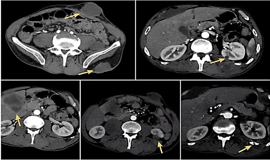
A computed tomography (CT) scan of the abdomen showed multiple solid lesions, namely a lesion located anterior to the rectum and posterior to the seminal vesicles (3.5cm) and an exophytic cortical lesion in the left kidney (3.2 cm), and other lesions on the V segment of the liver (5.4cm), right adrenal gland (3.3 cm), rectus abdominis muscle, and adjacent subcutaneous tissue (Fig. 4).

Considering the diagnosis of cutaneous metastases from a poorly differentiated urothelial carcinoma, possibly originating in the bladder and/or urinary tract, the patient started chemotherapy with carboplatin and paclitaxel but he died within 2 weeks.

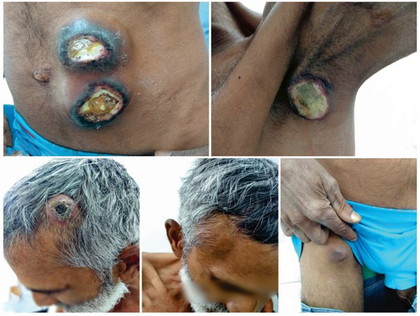
Figure 1 Ulcerated tumors in the abdominal region (A), left axilla (B), left frontotemporal region (C), large subcutaneous tumors in the right frontotemporal region (D) and right inguinal region (E). 

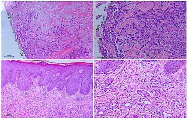
Figure 2 Histopathology of a skin biopsy showing dermal infiltration by polygonal neoplastic cells in A - (40× H&E), with more detail in B - (100×H&E), preservation of the epidermis in C - (40× H&E) and angiolymphatic invasion by nests of neoplastic c ells in D - (100× H&E). 

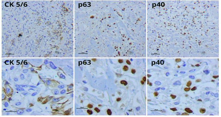
Figure 3 Immunohistochemistry with positive focal immunostaining for CK5/6, p63, and p40 (100× and 400× H&E). 

Figure 4 Abdominal CT scan showing multiple solid lesions in the left abdominal rectus muscle and adjacent subcutaneous tissue, in the ipsilateral gluteal muscle, in the hepatic segment, renal cortex, and a lin ear fracture of the left twelfth rib. 

DISCUSSION

Cutaneous metastases generally present as single or multiple nodules and tumors, with stony consistency; they may ulcerate and present a necrotic aspect, as observed in our patient.² Occasionally, they present as erythematous, sclerodermiform or papulonodular lesions.² Diagnosis can be difficult due to similarities with primary cutaneous lesions, such as hemangiomas, keratoacanthomas,3 sarcoidosis, leprosy, boils, sebaceous cysts, and opportunistic infections.4 Histopathology usually shows neoplastic cells that infiltrate the dermis and preserve the epidermis and may invade dermal vessels,5 which is also the pattern of urothelial carcinomas,6 as in the reported case. The immunohistochemistry profile with positivity for cytokeratins 5/6/7 and p63 is concordant with other cases in the literature, but cytokeratin 20 which, along with cytokeratin 7, is positive in 89% of transitional cell bladder cancers,7 was not positive in this case.

Urothelial neoplasms occur mostly in the bladder (90%), are approximately 3 times more frequent in men between the fifth and seventh decade. Main risk factors are tobacco smoke (which is also associated with higher disease spread through the urothelium to adjacent structures, as observed in the present case), occupational exposure to aromatic amines, cyclophosphamide, improper use of paracetamol, chronic irritation secondary to infected nephrolithiasis, and cystitis caused by Schistosoma haematobium.9,10,11

Symptoms related to urothelial neoplasia are scarce, the main one being macroscopic hematuria,2 which became apparent in this case only after cutaneous metastasis. Most patients are diagnosed during routine examination or due to metastatic lesions,12 that are mainly localized in the head, face, neck, trunk, abdomen, suprapubic region or extremities.7 Nevertheless, only approximately 20% of these cutaneous metastases occur at the initial presentation.8 They generally develop late in the evolution of the disease and cutaneous involvement indicates a poor prognosis.

In this case cutaneous lesions were the presenting sign of an urothelial carcinoma but they occurred only after disease dissemination and were, therefore, associated with a very poor prognosis. Recognition of cutaneous metastases can be

important to the diagnosis of internal malignancies, particularly in early stages, when treatment may have a favorable impact on prognosis.

Acknowledgments

The authors would like to thank Dr. Sofia Santos Tenório for contributing substantially to analysis of the histopathological material and for providing and describing the slide images.

REFERENCES

1. Azulay RD, Azulay L. Dermatologia. 7ª ed. São Paulo: Guanabara Koogan; 2017. [ Links ]

2. Soares RO, Correia TP, Lima P, Íscar-Galán T, Cardoso A, Reis FC, et al. Carcinoma urotelial do bacinete apresentando- se com metastização cutânea, gástrica e rectal. Acta Urol; 2010; 3: 75-8. [ Links ]

3. De Giorgi V, Sestini S, Massi D, Panelos J, Grazzini M, Lotti T. Cutaneous metastases: an ominous sign of urothelial carcinoma of the bladder. Dermatol Surg 2008; 34: 1574-6. doi: 10.1111/j.1524-4725.2008.34326.x. [ Links ]

4. Puerto JH, Flores JH, Casas JM, Velasco JJ. Metástasis cutánea de un carcinoma vesical de células transicionales. Actas Urol. Esp. 2002; 26: 413-5. doi: 10.1016/s0210-4806(02)72803-9. [ Links ]

5. Swick BL, Gordon JR. Superficially invasive transitional cell carcinoma of the bladder associated with distant cutaneous metastases. J Cutan Pathol. 2010; 37: 1245-50. doi: 10.1111/j.1600-0560.2009.01471.x. [ Links ]

6. Bernardes Filho F, Neves DG, Melo AS, Cruz MF, Pires AR, Kac BK, Lupi O. Invasão cutânea por carcinoma urotelial sarcomatoide: características clínicas e dermatopatológicas. An Bras Dermatol. 2016; 91: 73-9. [ Links ]

7. Lees AN. Cutaneous metastasis of transitional cell carcinoma of the urinary bladder eight years after the primary: a case report. J Med Case Rep. 2015; 9:102. doi: 10.1186/s13256-015-0585-9. [ Links ]

8. Messing EM. Urothelial tumors of the urinary tract. In: Walsh PC, Retik AB, Vaughan ED Jr, Wein AJ, Kavoussi LR, Novick AC, et al, editors. Campbell's urology. 8th ed. Philadelphia: Saunders; 2002. p. 2732-84. [ Links ]

9. Longo DL. Hematologia e Oncologia de Harrison. 2ºed. São Paulo: AMGH Editora Ltda; 2015. [ Links ]

10. Ribeiro DC, Amorim de Brito G, Oliveira CL, Fosse Júnior AM, Lima FV, Scheinkman J. Carcinoma Urotelial de Trato Urinário Superior Multifocal de Alto Grau. Relato de caso. Urominas. Rev Cient Urol SBU-MG; 2016; 3:48-51. [ Links ]

11. Lucon AM, Falci Junior R. Câncer de bexiga. In: Lopes AC, editor. Tratado de Clínica Médica. São Paulo: Roca; 2006. p. 2923-30. [ Links ]

12. Weiss L, Harlos JP, Torhorst J, Gunthard B, Hartveit F, Svendsen E, et al. Metastatic patterns of renal carcinoma: Na analysis of 687 necropsies. J Cancer Res Clin Oncol. 1988; 114:605-12. doi: 10.1007/BF00398185. [ Links ]

13. Block C, Dahmoush L, Konety B. Cutaneous metastases from transitional cell carcinoma of the bladder. Urology. 2006; 67:846.e15-7. doi: 10.1016/j.urology.2005.10.045. [ Links ]

14. Jakse G, Stockle M, Lehmann J, Otto T, Krege S, Rubben H. Metastatic Bladder Carcinoma. Dtsch Arztebl. 2007; 104:1024-8. [ Links ]

15. Salemis NS, Gakis C, Zografidis A, Gourgiotis S. Cutaneous metastasis of transitional cell bladder carcinoma: a rare presentation and literature review. J Cancer Res Ther. Apr-Jun 2011;7:217-9. doi: 10.4103/0973-1482.82940 [ Links ]

3© Autor (es) (ou seu (s) empregador (es)) 2020 Revista SPDV. Reutilização permitida de acordo com CC BY-NC. Nenhuma reutilização comercial. © Author(s) (or their employer(s)) 2020 SPDV Journal. Re-use permitted under CC BY-NC. No commercial re-use

Received: April 23, 2020; Accepted: July 18, 2020

Correspondência: Tânia Rita Moreno de Oliveira Fernandes Av. José de Sá Maniçoba, S/N - Centro 56304-917 - Petrolina - PE, Brazil E-mail:trmofernandes@gmail.com

Conflitos de interesse: Os autores declaram a inexistência de conflitos de interesse na realização do presente trabalho. Fontes de financiamento: Não existiram fontes externas de financiamento para a realização deste artigo. Confidencialidade dos dados: Os autores declaram ter seguido os protocolos da sua instituição acerca da publicação dos dados de doentes. Consentimento: Consentimento do doente para publicação obtido. Proveniência e revisão por pares: Não comissionado; revisão externa por pares

Conflicts of interest: The authors have no conflicts of interest to declare. Financing support: This work has not received any contribution, grant or scholarship. Confidentiality of data: The authors declare that they have followed the protocols of their work center on the publication of data from patients. Patient Consent: Consent for publication was obtained. Provenance and peer review: Not commissioned; externally peer reviewed

Creative Commons License This is an open-access article distributed under the terms of the Creative Commons Attribution License