SciELO - Scientific Electronic Library Online

 
vol.16 número4Impacto prognóstico dos biomarcadores inflamatórios pre-operatórios em contexto de isquemia aguda de membro: revisão sistemáticaExtensão proximal de trombose aórtica juxtarenal: uma complicação subestimada? índice de autoresíndice de assuntosPesquisa de artigos
Home Pagelista alfabética de periódicos  

Serviços Personalizados

Journal

Artigo

Indicadores

Links relacionados

  • Não possue artigos similaresSimilares em SciELO

Compartilhar


Angiologia e Cirurgia Vascular

versão impressa ISSN 1646-706X

Angiol Cir Vasc vol.16 no.4 Lisboa dez. 2020  Epub 31-Jan-2021


Caso clínico

Radial pulsatile mass - appearances can be deceiving

Massa radial pulsátil - as aparências iludem

Ana Carolina Semião1  2 

Clara Nogueira1  2 

Andreia Coelho1  2 

Ricardo Gouveia1  2 

Rita Augusto1  2 

Nuno Coelho1  2 

Evelise Pinto1 

João Ribeiro1  2 

João Peixoto1  2 

Luís Fernandes1 

Alexandra Canedo1  2 

1 Department of Angiology and Vascular Surgery, Centro Hospitalar de Vila Nova de Gaia/Espinho, Portugal

2 Faculty of Medicine of the University of Porto, Portugal.


Abstract

Adventitial cystic disease (ACD) is a rare condition. It affects mostly the popliteal artery, with few cases reported affecting the radial artery. This condition is characterized by accumulation of mucinous content at the adventitious layer.

Keywords: Adventitial cystic disease; Radial artery

Resumo

A doença quística da adventícia é uma condição rara. Afecta sobretudo a artéria poplítea, com poucos casos relatados que afectem a artéria radial. Esta doença é caracterizada pela acumulação de um conteúdo mucinoso na camada adventícia.

Palavras-chave: Doença quística da adventícia; Artéria radial

Case report

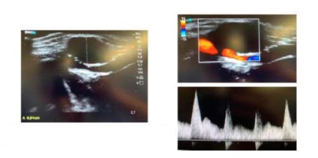
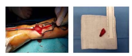
We report the case of a 44 year old healthy male, presenting with a 2 cm pulsatile, painless mass in the distal portion of the radial side of the forearm on the left wrist, with no previous history of trauma or percutaneous procedures. Doppler ultrasound (DUS) revealed a hypoechogenic mass involving the arterial wall that caused a lumen reduction of 50%. Lesions in other arterial segments were excluded using DUS. These findings were compatible with the diagnosis of ACD and patient was elective for surgery. The involved arterial segment was resected and an end-to-end anastomosis was performed. Pathology confirmed the presence of a cystic mass with mucinous content. During follow-up, the patient remained asymptomatic and DUS showed patency of the artery and no signs of recurrence.

Discussion

This rare entity should be suspected when a pulsatile mass is noted in the radial artery because an early diagnosis and appropriate management may prevent further complications.

ACD is characterized by the collection of mucinous material which accumulates in the adventitial layer of the vessels. The first case of this rare entity was reported in 1947, which involved the external iliac artery.(1) However, it affects predominantely the popliteal artery.(2) ACD may also involve other peripheral vessels, as the radial artery, but the percentage of reported cases in this location is limited to 2,5% of all ACD cases.(3)

To date, its etiology still has not been clarified. Several hypotheses have been proposed to explain its formation: cystic degeneration of the adventitia as a consequence of repetitive trauma(4,5); adventitial degeneration due to a systemic process; inclusion of mesenchymal cells within the adventitia(6,7,8); and development from a direct communication with an adjacent joint space(8).

ACD affects males predominantly, with a male to female ratio of 5 to 1, and patients are typically in their mid-40s. Some investigators have reported a slightly older age at diagnosis(9).

Pathology reveals that these cysts are filled with gelatinous mucoid material. A closer microscopic examination reveals a cuboid cell lining in the adventitial layer; atherosclerotic disease is absent.

The most common clinical presentation of ACD in the radial artery is as a newly detected asymptomatic pulsatile arterial mass. For this reason, the diagnosis of radial artery aneurysm/pseudoaneurysm should be excluded.

Apparently, the enlargement of the cyst is slow but progressive compression of the lumen can lead to arterial thrombosis with ischemic symptoms. In the radial artery, compression of nerve fibers has also been described, which may lead to presentation of paresthesias, pain, weakness or even thenar atrophy(10,11).

Diagnostic workup can involve angio-CT but current recommendations support DUS followed by MR or CT(12). DUS allows the evaluation of the cyst dimensions and the hemodynamic caractheristics of the vessel. Regarding diagnosis, these cysts should be differenciated from lipomas, fibromas or aneurysms. During follow-up, DUS is considered the first-line imaging for surveillance.

Treatment options are variable: nonsurgical management, cyst aspiration (percutaneous or open), angioplasty or surgical treatment. Surgical options include cyst incision and decompression, cyst resection alone or with repair with a patch (venous or synthetic) or reconstruction with a graft (venous or synthetic). The most common surgical option has been cyst resection and reconstruction with a reversed segment saphenous vein autograft(12,13).

When cystic ligation of the joint connection is performed as part of treatment, cyst recurrence has not been identified. On the other hand, leaving the articular branch has been related to failure of the procedure, leading to cyst reaccumulation. Recurrence or persistence rate of 9% has been reported, most of them occurred after less invasive techniques such as angioplasty or percutaneous aspiration(3).

In conclusion, although extremely uncommon, ACD of the radial artery should be suspected when a pulsatile mass is noted. An early diagnosis and appropriate management may prevent further complications.

Fig. 1 DUS with flow in the radial artery and anecogenc mass corresponding to the adventicial cyst. 

Fig. 2 Isolation of the arterial artery with exposure of the cyst (left). Adventicial cyst after being removed (right). 

REFERENCES

1. Atkins HJ, Key JA. A case of myxomatous tumour arising in the adventitia of the left external iliac artery; case report. Br J Surg. 1947;34(136):426. [ Links ]

2. Augustiny N, Goebel N, Jager K, Brunner U, von Schulthess GK. Cystic adventitial degeneration of the popliteal artery: nuclear magnetic resonance tomography and computerized tomography. Rofo. 1988;148(4):452-3. [ Links ]

3. Desy NM, Spinner RJ. The etiology and management of cystic adventitial disease. J Vasc Surg. 2014;60(1):235-45, 245.e1-11 [ Links ]

4. Flanigan DP, Burnham SJ, Goodreau JJ, Bergan JJ. Summary of cases of adventitial cystic disease of the popliteal artery. Ann Surg. 1979;189(2):165-75. [ Links ]

5. Levien LJ, Benn CA. Adventitial cystic disease: a unifying hypothesis. J Vasc Surg. 1998;28(2):193-205. [ Links ]

6. Maged IM, Turba UC, Housseini AM, Kern JA, Kron IL, Hagspiel KD. High spatial resolution magnetic resonance imaging of cystic adventitial disease of the popliteal artery. J Vasc Surg. 2010;51(2):471-4. [ Links ]

7. Ohta T, Kato R, Kondo M, Tsuchioka H. Recurrence of cystic adventitial disease in an interposed vein graft. Surgery. 1994;116(3):587-92. [ Links ]

8. Flessenkaemper I, Muller KM. Early recurrence of cystic adventitial disease in a vein graft after complete resection of the popliteal artery. Vasa. 2014;43(1):69-72. [ Links ]

9. AbuRahma AF et al. Rutherford's vascular surgery and endovascular therapy, nineth edition.2019. [ Links ]

10. Wang H, Spinner RJ, Amrami KK. Adventitial cyst of the radial artery with a wrist joint connection. J Hand Surg Am. 2007;32(1):126-30. [ Links ]

11. Durham JR, McIntyre KE Jr. Adventitial cystic disease of the radial artery. J Cardiovasc Surg (Torino). 1989;30(3):517-20. [ Links ]

12. Vanhoenacker FM, Vandevenne JE, De Schepper AM, De Leersnijder J. Regarding »,» ®,® §,§ ­,­ ¹,¹ ²,² ³,³ ß,ß Þ,Þ þ,þ ×,× Ú,Ú ú,ú Û,Û û,û Ù,Ù ù,ù ¨,¨ Ü,Ü ü,ü Ý,Ý ý,ý ¥,¥ ÿ,ÿ ¶,¶ Adventitial cystic disease: a unifying hypothesis »,» ®,® §,§ ­,­ ¹,¹ ²,² ³,³ ß,ß Þ,Þ þ,þ ×,× Ú,Ú ú,ú Û,Û û,û Ù,Ù ù,ù ¨,¨ Ü,Ü ü,ü Ý,Ý ý,ý ¥,¥ ÿ,ÿ ¶,¶ . J Vasc Surg. 2000;31(3):621-2. [ Links ]

13. Baxter AR, Garg K, Lamparello PJ, Mussa FF, Cayne NS, Berland T. Cystic adventitial disease of the popliteal artery: is there a consensus in management? Vascular. 2011;19(3):163-6. [ Links ]

14. Hernandez Mateo MM, Serrano Hernando FJ, Martinez Lopez I, Gonzalez Sanchez S, Hernando Rydings M, Saiz Jerez A, Revuelta Suero S, Marques de Marino P. Cystic adventitial degeneration of the popliteal artery: report on 3 cases and review of the literature. Ann Vasc Surg. 2014;28(4):1062-9. [ Links ]

Received: August 26, 2020; Accepted: December 28, 2020

Autor para correspondência. Correio eletrónico: anasemiao1@gmail.com (A. Semião).

Creative Commons License This is an open-access article distributed under the terms of the Creative Commons Attribution License