SciELO - Scientific Electronic Library Online

 
vol.16 número4Evolução da formação em cirurgia vascular nos últimos 15 anos em PortugalAneurismas tóraco-abdominais após dissecção aórtica: Resultados do tratamento aberto e endovascular índice de autoresíndice de assuntosPesquisa de artigos
Home Pagelista alfabética de periódicos  

Serviços Personalizados

Journal

Artigo

Indicadores

Links relacionados

  • Não possue artigos similaresSimilares em SciELO

Compartilhar


Angiologia e Cirurgia Vascular

versão impressa ISSN 1646-706X

Angiol Cir Vasc vol.16 no.4 Lisboa dez. 2020  Epub 31-Jan-2021


Artigo original

False aneurysms of the Renal Artery: is it a known diagnosis in the medical community?

Falsos aneurismas da Artéria Renal: é este um diagnóstico conhecido na comunidade médica?

Marta Machado1 

Rui Machado1  2 

Daniel Mendes2 

Rui de Almeida1  2 

1 . Instituto de Ciências Biomédicas Abel Salazar - Universidade do Porto; Portugal

2 . Serviço de Angiologia e Cirurgia Vascular, Centro Hospitalar Universitário do Porto. Portugal


Abstract

Introduction:

False aneurysms of the Renal Artery usually originate from iatrogenic causes or renal trauma. The incidence is increasing because minimally invasive procedures have become widely used in the treatment of renal pathology and therefore iatrogenic injuries have become more frequent.

Due to the nature of the wall, they have a high risk of rupture and should therefore be diagnosed and treated soon.

Objective:

To evaluate the experience of the CHUP’s angiology and vascular surgery department in the approach of false aneurysms of the renal artery from 2010 to 2018.

Methods and Material:

A retrospective review was undertaken using the clinical and imagiological records of 20 patients diagnosed with FRA.

Results: Etiology was iatrogenic in 95% of the cases. All patients were symptomatic and 85% had macroscopic hematuria. The time between injury and clinical manifestation was 4.5 days. The therapy performed in all cases consisted of embolization of the FRA feeding artery. The time between injury and treatment was 18.1 days. The kidney salvage rate was 95%.

Discussion/Conclusion:

In this study, the main etiology and the surgical technique used are in agreement with the published literature. With this kidney salvage rate we can conclude that endovascular treatment is an effective method of treatment of these lesions.

Although there are no published references in the literature, we observed that the time between injury and treatment was high, which translates into a late diagnosis. Therefore, the urologists' awareness of this complication is extremely important.

Keywords: Renal artery aneurysms; False aneurysms; Pseudoaneurysms; Endovascular treatment

Resumo

Introdução:

Os falsos aneurismas da artéria renal são maioritariamente causados por lesões iatrogénicas ou por trauma renal. A incidência destes está a aumentar devido ao incremento do uso de procedimentos minimamente invasivos no tratamento da patologia renal e, consequente aumento das lesões iatrogénicas. Devido à natureza da parede, estes apresentam um alto risco de rutura e, portanto, devem ser diagnosticados e tratados precocemente.

Objetivo:

Avaliar a experiência do Departamento de Angiologia e Cirurgia Vascular do CHUP na abordagem dos falsos aneurismas da artéria renal.

Métodos e materiais:

Foi realizada uma revisão retrospetiva com recurso aos registos clínicos e imagiológicos de 20 pacientes com diagnóstico de falsos aneurismas da artéria renal entre 2010 a 2018.

Resultados:

Em 95% dos casos a etiologia foi iatrogénica. Todos os pacientes apresentaram-se sintomáticos, 85% com hematúria macroscópica. O tempo médio entre a lesão e a manifestação clínica foi de 4,5 dias. O tratamento realizado em todos os casos consistiu na embolização da artéria alimentadora. O tempo entre a lesão e o tratamento foi de 18,1 dias. A taxa de salvamento do rim foi de 95%.

Discussão/Conclusão:

Neste estudo, a principal etiologia e a técnica cirúrgica utilizada estão de acordo com a literatura publicada. Esta taxa de salvamento do rim, reforça que o tratamento endovascular é um método eficaz. Embora não haja referências publicadas na literatura que permitam a comparação, neste estudo observou-se que o tempo entre a lesão e o tratamento foi elevado, o que se traduz num diagnóstico tardio. Neste sentido, a consciencialização dos urologistas em relação a esta complicação é extremamente importante.

Palavras-chave: Aneurismas da artéria renal; Falsos aneurismas; Pseudoaneurismas; Tratamento endovascular

Introduction

False aneurysms of the Renal Artery (FRAs) usually originates from renal trauma or iatrogenic causes, such as vascular procedures (angioplasty and renal stenting), urological catheterizations or percutaneous renal procedures (biopsies, nephrostomies, nephroureterolithotomies and partial nephrectomy).(1-6)

The diagnosis of FRA can be incidental or due to hematuria, low back pain, abdominal pain, dizziness, syncope, hypotension or hypovolemic shock. The worsening of renal function is rare and is more frequent when there's a single kidney. The possible causes are the presence of blood clots in the excretory system (acute post-renal kidney injury) or the combination of hypotension and hypovolemia (acute pre-renal kidney injury).(7)

Due to the nature of the FRA wall, they have a high risk of rupture and should therefore be diagnosed and treated soon.(8)

The therapeutic options in the FRA are varied and related to the size of the FRA and clinical manifestations, ranging from the injection of echo-guided thrombin to treatment by endovascular or open surgery. Open surgery usually consists in nephrectomy because in most cases the vessels that are involved are small intra-parenchymal vessels and so the endovascular treatment is the only that can minimize the loss of kidney tissue. Recent developments in endovascular surgery have made embolization by superselective catheterization of the artery involved the treatment of choice. The rate of endovascular complications is low, with the rate of occlusion of the main renal arteria and consequent renal infarction being minimal.(9-11)

Objectives

To evaluate the experience of the CHUP’s angiology and vascular surgery department in the approach of renal artery pseudoaneurysms.

Methods and material

A retrospective review was undertaken using the clinical and imagiological records of 20 patients diagnosed with renal artery pseudoaneurysm from 2010 to 2018. SPSS version 21 was used for the treatment and analysis of the descriptive statistics (univariate and bivariate) , hypothesis tests and analysis of probability of accumulated survival.

Results

60% of the patients were men and the average age was 55.7 + -19.2 years, with at least 6 to 86 years old.

The etiological cause of FRA in 95% of the cases was iatrogenic due to an urological / nephrological procedure (35% due to open partial nephrectomy (OPN), 25% due to biopsies, 20% due to laparoscopic partial nephrectomy (LPN), 15% due to percutaneous procedures) and one case due to trauma.

The diagnosis was based on clinical suspicion (85% had macroscopic hematuria) and all the cases were confirmed with CT. In 30% of the cases, an association with AVF was observed.

The therapy performed in all cases consisted of embolization of the FRA feeding artery, with embolization with coils being the most used method (65% in total - 30% microcoils + 35% coils) (Table I)

Table I Descriptive analysis 

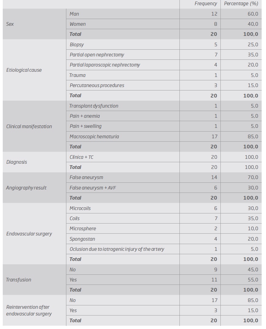
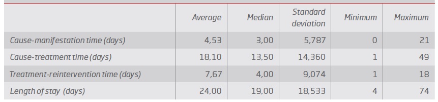
The time between the lesion and the clinical manifestation was 4.5 days, the time between the lesion and the treatment was 18.1 days. (Table II)

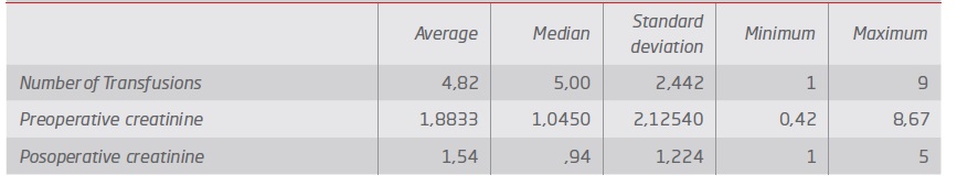
The transfusion rate was 55% with an average of 4.8 units of red blood cells and an average of length of stay of 24 days.

There was no statistically significant difference between pre and postoperative creatinine. (Table II and III).

Mortality was 0%, the rebleeding rate (which occurred in the first two weeks) requiring a second embolization was 15% and the kidney salvage rate was 95% with an average follow-up time of 96 weeks. (Chart 1)

Chart 1 Endovascular reintervention curve 

Discussion/ Conclusion

In our series, the average age at diagnosis was 55.7 years with a predominance of males (60%). Guo et al(12) reported an average age of 46.6 years and a male involvement of 63%.

Partial nephrectomy (PN) was the most common etiological cause, 35% for OPN (open partial nephrectomy) and 20% for LPN (laparoscopic partial nephrectomy) (Figures 1 e 2). In the study by Guo et al(12), LPN represented 25.9% of the etiological causes. Zhu Y et al(13) published an incidence of FRA after LPN (4.26%) higher than after OPN ( 3.17%). In fact, in our study, we cannot calculate this rate but we believe that the reason why the percentage of FRA after OPN is higher is because the study is made with records between 2010-2018 and therefore the absolute total number of OPN done in this period is also much higher than absolute total number of LPN done in the same period. Renal biopsies were the etiological cause in 25% of our cases (Figure 3). In the study by Guo et al(12) renal biopsies represent 51.9% of the etiological causes, a value much higher than that of our series.

Percutaneous urological procedures (in our case, percutaneous nephrolithotomy-NLP) represented 15% of the etiological causes. In the study by Guo et al(12) this was the cause in 14.8% of FAR. Poyraz N et al(16) described 14 cases of FAR and 5 of AVF in 1609 patients undergoing NLP, with all patients presenting macroscopic hematuria or massive hematuria and all requiring blood transfusion. Coil embolization was the preferred treatment, with only two patients developing recurrence of hemorrhage and treated for further embolization.

Table II Time between cause, diagnosis, treatment and length of stay 

Table III Need for red cells transfusion and repercussion on the kidney function 

In only one case (5%), the etiology was blunt kidney trauma (Figure 4). According to Ierardi AM et al(17) kidney injuries occur in approximately 1-5% of all trauma patients.

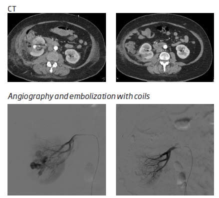
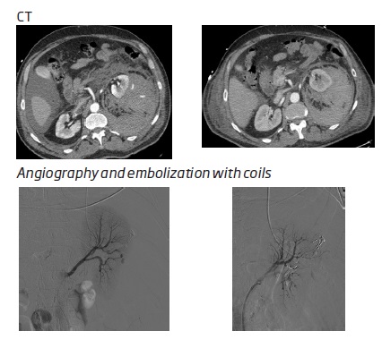
Figure 1 False right renal aneurysm, after conventional partial nephrectomy treated by embolization with coils 

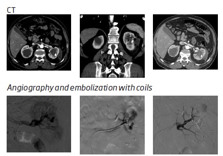
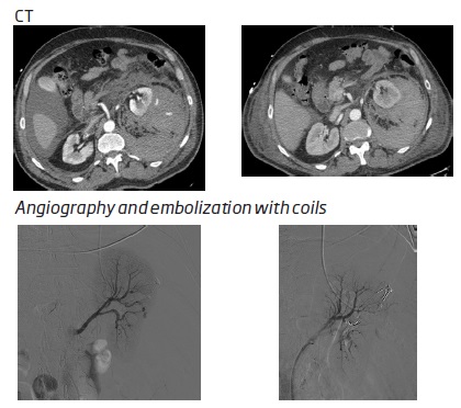
Figure 2 False left renal aneurysm with associated VAF, after laparoscopic segmental nephrectomy treated with embolization with coil 

Figure 3 False aneurysm after biopsy of native kidney treated with embolization with coils 

Figure 4 False left renal aneurysm, after blunt trauma treated by embolization with coils 

Endovascular surgery is currently the first therapeutic line that can prevent nephrectomy by up to 78%-83%,(18) although traditionally high-grade kidney injuries are corrected by conventional surgery.

The most common clinical manifestation of FAR was macroscopic hematuria that occurred in 85% of patients. According to the literature, 11.2 to 17.5%(19) of hemorrhages endanger the patient's life. The diagnosis of FAR in all cases was based on clinical suspicion and was confirmed by CT. According to Guo et al(12), the first exam to be performed in the clinical suspicion of FAR should be ultrasound, to avoid the contrast overload associated with CT. However, we believe that the CT it will always be necessary because it allows for diagnosis and surgical planning and is independent of the patient's biotype and of the technician who performs it.

Pitton M B et al(2) reported a rupture rate of 76.3% in FAR compared to 3.1% in true aneurysms, and there do not seem to be any predictive factors for rupture. Therefore, the literature argues that FRAs should be treated after being diagnosed, which is in line with our series in which all FARs were treated surgically.

In our series, the therapy performed consisted of embolization of the FAR feeding artery in all cases, with embolization with coils being the most used method. Mortality was 0%, the bleeding recurrence rate requiring a second embolization was 15% and the kidney salvage rate was 95%. There was no statistically significant association between the etiology of FAR and the number of transfusions and therapeutic reintervention. Ghoneim et al(22) reported that the success rate was 90 to 95% success(23,24) and that selective renal embolization does not cause obvious changes in the glomerular filtration rate, which is in accordance with our results (mean preoperative creatinine 1.9 and 1.5 with no no statistically significant difference).

The time between the injury and the clinical manifestation was 4.5 days, the time between the injury and the treatment was 18.1 days. The transfusion rate was 55% and the average length of stay was 24 days. Hongjie G et al reported lower values for the time between injury and treatment (3 days), with 88.9% of patients treated in the first 14 days. These long times and the need to carry out a large number of transfusions, make us suppose that a better knowledge of this pathology can improve these results. The urologist must be familiar with this clinical entity to make a quick diagnosis and promote an efficient treatment.

References

1. Santucci, RA, et al. Validation of the American Association for the Surgery of Trauma organ injury severity scale for the kidney. J Trauma. 2001; 50:195-200. [ Links ]

2. Mani, NB e Kim, L. The role of interventional radiology in urologic tract trauma. Semin Intervent Radiol. 2011; 28:415-23. [ Links ]

3. Loffroy, R, et al. Role of percutaneous arterial embolization in renal pathology. Prog Urol. 2010; 20:161-71. [ Links ]

4. Bittenbinder, EN e Reed, AB. Advances in renal intervention for trauma. Semin Vasc Surg. 2013; 26:165-9. [ Links ]

5. Loffroy, R, et al. Transcatheter arterial embolization in patients with kidney diseases: an overview of the technical aspects and clinical indications. Korean J Radiol. 2010; 11:257-68. [ Links ]

6. Loffroy, R, et al. Management of post-biopsy renal allograft arteriovenous fistulas with selective arterial embolization: immediate and long-term outcomes. Clin Radiol. 2008;63:657-65. [ Links ]

7. Antunes,I, et al. False aneurysms of the renal artery our experience. Angiol Cir Vasc. 2015; 11:199-203 [ Links ]

8. Pitton, MB, et al. Visceral artery aneurysms: incidence, management, and outcome analysis in a tertiary care center over one decade. Eur Radiol. 2015; 25:2004-14. [ Links ]

9. Zabkowski, T, et al. Superselective renal artery embolization in the treatment of iatrogenic bleeding into the urinary tract. Med Sci Monit. 2015;21:333-7. [ Links ]

10. Poulakis, V, et al. Treatment of renal-vascular injury by transcatheter embolization: immediate and long-term effects on renal function. J Endourol. 2006; 20:405-9. [ Links ]

11. Schwartz, MJ, et al. Renal artery embolization: clinical indications and experience from over 100 cases. BJU Int. 2007; 99:881-6. [ Links ]

12. Guo, H, et al. Management of iatrogenic renal arteriovenous fistula and renal arterial pseusdoaneurysm by transarterial embolization - a single center analysis and outcomes. Medicine. 2017;40(96):1-5. [ Links ]

13. Zhu, Y, et al. Risk evaluation of selective renal arterial embolization in the treatment of delayed hemorrhage from renal artery pseudoaneurysm after partial nephrectomy. Cell Biochem Biophys. 2014; 3(70):1833-9. [ Links ]

14. Brandenburg, VM, Frank, RD e Riehl, J. Color-coded duplex sonography study of arteriovenous fistulae and pseudoaneurysm complicating percutaneous renal allograft biopsy. Clin Nephrol. 2002;58:398-404. [ Links ]

15. Preda, A, et al. Complication rate and diagnostic yield of 515 consecutive ultrasound-guided biopsies of renal allografts and native kidneys using a 14-gauge Biopty gun. Eur Radiol. 2003; 13:527-30. [ Links ]

16. Poyraz, N, et al. Clinical efficacy and safety of transcatheter embolization for vascular complications after percutaneous nephrolithotomy. Videosurgery and other miniinvasive techniques. 2017; 4:403-8. [ Links ]

17. Ierardi, AM, et al. Transcatheter of iatrogenic renal vascular injuries. Radiol Med. 2014; 119:261-8. [ Links ]

18. Ramaswamy, RS e Darcy, MD. Arterial Embolization for the Treatment of Renal Masses and Traumatic Renal Injuries. Tech Vasc Interv Radiol. 2016; 19:203-10. [ Links ]

19. Caliskan, KC, et al. How and when to perform transcatheter embolization in iatrogenic renal hemorrhage? A new algorithm. Med J Okmeydani Train Res Hosp. 2012;28:84-93. [ Links ]

20. Ngo, TC, Lee, JJ e Gonzalgo, ML. Renal pseudoaneurysm: an overview. Nat Rev Urol. 2010;7:619-25. [ Links ]

21. Rivera, M, et al. Asymmptomatic large extracapsular renal pseudoaneurysm following kidney transplant biopsy. Am J Kidney dis. 2011; 57:175-178. [ Links ]

22. Ghoneim, TP, et al. Selective arterial embolization for pseudoaneuryms and arteriavenous fistula of renal arterybranches following partial nephrectomy. Journal of Urology. 2016; 185. [ Links ]

23. Shapiro, EY, et al. Renal artery pseudoaneurysm following laparoscopic partial nephrectomy. Urology. 2009; 74:819. [ Links ]

24. Singh, D e Gill, IS. Renal artery pseudoaneurysm following laparoscopic partial nephrectomy. Journal of Urology. 2005; 174:2256. [ Links ]

Received: October 06, 2020; Accepted: December 30, 2020

Autor para correspondência. Correio eletrónico: marta_machado_vb@hotmail.com (M. Machado).

Creative Commons License This is an open-access article distributed under the terms of the Creative Commons Attribution License