


 English (pdf)
 Article in xml format
 Article references
 Send this article by e-mail
 Cited by SciELO
  Similars in
SciELO
Similars in
SciELO 
 Permalink
Introduction
Erythema multiforme (EM) is a disease characterized by na acute mucocutaneous hypersensitivity reaction, with typical ulcerative skin eruptions in the form of erythematous concentric rings, called target-type lesions, that may involve the oral cavity or other mucosa.1,2 Although its etiopathogenesis is still uncertain, it has been suggested that EM may be induced by the ingestion of some drugs, such as antibiotics, analgesics, anticonvulsants, non-steroidal anti-inflammatory drugs, and antifungals, or by infections, particularly by the herpes simplex virus (HSV) and the bacterium Mycoplasma pneumoniae, which is commonly reported, especially in children.1,3,4
According to the number of mucosal sites involved and the severity of the lesions, the disease can be subclassified as minor or major EM.2 Minor EM is the mildest form of the disease, with target-type lesions affecting less than 10% of the body surface area and only one mucosa, usually the mouth.1,5 Stevens-Johnson syndrome (SJS), a severe variant of EM, usually has more generalized skin involvement and prodromal, flu-like systemic symptoms. Toxic epidermal necrolysis is even more severe, characterized by epidermal detachment from the body surface and purpuric macules or generalized atypical flat target lesions.1,2,5
Although skin lesions are important for diagnosing EM, ulcerations in the oral cavity without skin lesions have been reported, and some authors refer to this condition as oral EM.2,6,7 Considering its unusual manifestation, this work reports a case of EM restricted to the oral cavity and its treatment.
Case report
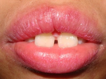
A 12-year-old female child complained of lesions in the vermilion of the lip and perioral skin with edema and bleeding, with minimal manipulation. The patient had extremely painful symptoms, which made speaking, chewing, and thorough intraoral examination difficult. The patient also exhibited a history of recurrence. Upon clinical examination, extensive ulcerative lesions were observed on the upper and lower lips, extending to the perioral region and the dermis, with bloody, suppurative, and crusted areas (Figure 1). The patient did not report any febrile episodes or medication consumption prior to the onset of these lesions. No other ulcerative or vesiculobullous changes were observed in other parts of the body, and there was no involvement of other mucous membranes (Figure 2). Based on the symptoms, the condition was diagnosed as EM, despite the absence of other skin eruptions, with lesions restricted to the lip region. Pharmacological treatment was recommended, with 4 mg of dexamethasone prescribed twice a day for seven days, in addition to the use of a medicated lip balm (clobetasol).
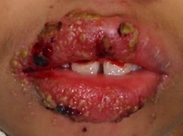
Figure 1 Clinical symptoms of oral erythema multiforme (EM) at the first visit of the patient. Manifestation of ulcerative lesions on the upper and lower lip regions
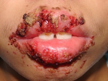
One week later, there was no significant regression of the condition, with persisting extensive ulcerative hemorrhagic lesions and painful symptoms (Figure 3). Owing to the persistence of infection in the lesions (suppurative crusts), the pharmacological treatment was modified by prescription of another corticosteroid and an antibiotic, as follows: prednisone, 10 mg, twice a day, for seven days; 500 mg of amoxicillin, thrice a day, for seven days. Use of a 0.12% chlorhexidine-gluconate antiseptic mouthwash was indicated to improve oral hygiene, which was affected due to the pain and difficulty in opening the mouth.
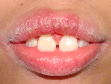
In the second return evaluation, after the last pharmacological prescription ended, considerable regression of the lesions and lip dryness was observed, with some small ulcers already in the healing process (Figure 4). In addition to wide mouth opening, the patient was able to speak and eat with more ease. In the third visit, the lesions had disappeared entirely, and only lip dryness was observed (Figure 5), so the patient was advised to continue using lip balm. Follow-up was performed for one year, and no recurrence of EM was observed in the patient.
Discussion and conclusions
EM is an acute mucocutaneous condition that is sometimes recurrent and usually self-limiting. Its main triggers include two broad categories: infection (particularly by HSV) and drugs, such as antibiotics, analgesics, anticonvulsants, non-steroidal anti-inflammatory drugs, and antifungals.1,2
The host responds to the external antigen via an immune-mediated reaction involving T cells, leading to a cytotoxic immune attack that results in keratinocyte lysis.4,5Other ocasional factors include malignancies, autoimmune, genetic factors, chemicals, and radiation therapy. Vaccines such as those against measles, mumps and rubella, smallpox, meningococcal hepatitis B, pneumococcal, chickenpox, influenza, Haemophilus influenzae, and even COVID-19 have been linked to EM, although with a low incidence. Despite knowledge of the existence of a cell-mediated immune response, its exact etiopathogenesis remains uncertain.2,5,8
In the present case, the patient did not report having previous episodes of HSV infection or having taken any medication. As no additional tests were performed to detect HSV, its role in the development of the lesions cannot be excluded. Furthermore, HSV-associated EM can develop without clinical manifestations of herpetic viral infection.4 In the 1-year follow- up period, the patient did not develop disease recurrence.
Relapse of EM is generally secondary to the reactivation of HSV; thus, the virus may be present in patients with an apparent idiopathic recurrence of EM and no clinical history of HSV infection or drug intake.9
Diagnosis of EM is primarily clinical, facilitated by the history of acute onset of mucocutaneous lesions, and may be preceded by the aforementioned factors combined with the appearance of typical target-type lesions.4 In minor EM, the disease lesions are characterized by single mucosal ulcerations and distinctive target-type lesions on the skin. In major EM, a more aggressive form, multiple mucosae are involved in addition to the typical target-type lesions.5 Some conditions should be considered as a differential diagnosis for EM, such as pityriasis rosea, urticaria, viral exanthema, fixed drug eruption, bullous pemphigoid, SJS, polymorphic light eruption, paraneoplastic pemphigus, and hypersensitivity reactions. The differential diagnosis should consider the following typical symptoms of EM: lesions lasting a minimum of seven days, as opposed to individual urticarial lesions that usually resolve within a day; more widespread lesions, while fixed drug eruptions commonly exhibit fewer lesions; and formation of papular patterns on the skin, as opposed to the disseminated erythematous or purpuric macules with blisters typical of SJS.10 In the present case, the patient had the lesions for more than 14 days, several ulcerations in the upper and lower lips and perioral skin, and no previous related drug intake, corroborating the diagnosis of minor EM.
Lesion biopsy is unnecessary when the clinical picture is clear, as histopathological findings are not pathognomonic.4 Histopathological features may include intercellular and intracelular edema of the lining epithelium, focal formation of microvesicles, exocytosis, diffuse inflammatory infiltrate, vasodilation, and edema of the connective tissue.5 Direct immunofluorescence can help differentiate between EM and bullous autoimmune diseases, such as bullous pemphigoid.10
In the present case, the diagnosis of EM was made based only on clinical findings, without performing a biopsy, even in the absence of skin lesions. There have been reports of EM with lesions confined to the oral mucosa and lips, without compromising the skin in acral distribution, that comprises a third category called oral EM, consistent with the presente case findings.2,6,7
Treatment of EM varies according to disease severity and involves identifying the cause of the disease when possible.
Affected patients should discontinue all causal drugs and treat relevant infections as needed.4 Because oral EM can be very painful, the patient may reduce their food intake in more severe situations, which might lead to hospitalization for intravenous fluids and electrolyte replacement. Symptom management involves topical corticosteroids and short-term systemic prednisone, which has been reported to be eficiente in controlling oral EM lesions, as was in our patient.10 Anti-inflammatory drugs, analgesics, or topical anesthetics for symptomatic relief can also be used, as well as antiseptic solutions to aid oral hygiene.5 In cases of EM associated with HSV, control with acyclovir may be effective if its use starts in the first days of active viral infection.1 However, the manifestation of the disease may only occur eight days after infection development, at which point the treatment directed to the HSV infection is no longer indicated. Continuous antiviral therapy with acyclovir, valacyclovir, or famciclovir for six months (depending on the case) can be effective in preventing the recurrence of EM by HSV.5
Mouth-restricted EM is rare and may represent a challenging diagnosis because even if EM attacks are confined to the oral mucosa, disease recurrence can manifest more aggressively with skin involvement.2 Therefore, it is important to carry out a detailed anamnesis and physical examination to exclude other inflammatory or immunological diseases, such as lichen planus, pemphigus vulgaris, and herpetic lesions.7
Moreover, it is important to identify signs and symptoms of ulcerative disorders involving the oral cavity so that there is an adequate diagnosis, early treatment, and follow-up, where clinicians should keep EM as a possible diagnosis.

