


 Inglês (pdf)
 Artigo em XML
 Referências do artigo
 Enviar este artigo por email
 Citado por SciELO
  Similares em
SciELO
Similares em
SciELO 
 Permalink
Introduction
Dental anomalies of number can be defined as a change to the normal number of teeth present in the oral cavity. There are several clinical forms, such as anodontia (the complete absence of teeth), oligodontia (agenesis of six or more teeth, excluding the third molars), hypodontia (agenesis of up to six teeth, excluding the third molars), and hyperdontia (any case presenting supernumerary teeth).1,2
Hypodontia is the most common anomaly of number and the most frequent dentofacial anomaly. There are no diferences regarding the prevalence of primary tooth agenesis according to sex, but evidence suggests that permanent tooth agenesis is more common among females.1,2
While the prevalence of hypodontia sits somewhere between 1.6% and 36.5% for permanent teeth (deciduous tooth agenesis is considered much rarer, at under 1%),1,2 the prevalence of hyperdontia seems to be around 0.04% to 3%. Supernumerary teeth are more common among males.3-5They can be classified according to the region of the oral cavity in which they are found: mesiodens (in between the maxillary central incisors), paramolars (buccally or lingually to a molar), and distomolars (distally to the third molar).6-8
Hyperdontia is often associated with tooth impaction, rotation of teeth adjacent to the supernumerary tooth, periapical resorption (due to the development of a dentigerous cyst alongside the supernumerary tooth), and, in the case of the mesiodens, midline diastema. Hyperdontia has also been associated with several genetic entities and syndromes, such as cleidocranial dysplasia, familial adenomatous polyposis, and oculofaciocardiodental syndrome.3-5,8 Likewise, hypodontia has been associated with some craniofacial syndromes, including cleft lip, cleft palate, and Down’s syndrome. It can also be found without any associated genetic syndrome. Hypodontia is often accompanied by other dental anomalies, such as microdontia, delayed tooth development, and ectopic eruption of permanent teeth.2
Recently, some authors have studied the possibility of a link between tooth agenesis and some cancers.9-11 Genes connected with odontogenesis (and its failure) have been linked with some malignant tumors, namely: AXIN2, ATF1, DUSP10, CASC8 (all associated with colorectal cancer), and PAX9 (associated with malignant tumors of the esophagus and the ovaries).9,10 Despite the consensus that the evidence currently available for this association is limited, clinicians are advised to consider tooth agenesis as a potential early risk indicator for cancer, especially in cases where several teeth are congenitally absent. Still, dentists should be careful in choosing whether to communicate this information to their patients, opting to inform the patient only in the context of a multidisciplinar treatment plan involving an oncological medical professional or team.9,11
While diagnosing a case of tooth agenesis, one should be mindful of the chronology of eruption for both dentitions (deciduous and permanent). The last deciduous teeth to complete development are the upper canines, as their roots reach the final point of development around the age of 3,25 years old.12
Therefore, between the ages of 4 and 6 (when the primary teeth begin to exfoliate), the absence of a deciduous tooth indicates either its agenesis or its early extraction. At 4 years old, one could also expect to find radiographic signs of hard tissue formation for every permanent tooth, except for the third molars, as their hard tissue only begins forming between 7 and 9 years old (in the maxillary arch) or between 8 and 10 years old (in the mandibular arch).13
This study aimed to determine the prevalence of tooth agenesis and supernumerary teeth, and to characterize both anomalies, in a population of Portuguese pediatric patients attending the pedagogic clinic of the Faculty of Dental Medicine of the University of Porto (FMDUP). It focused on the association between sex and each of these dental anomalies, as well as the association between tooth agenesis and dental arch. The following null hypotheses were defined: “The variable ‘Diagnosis of Tooth Agenesis’ is independente of the variable ‘Sex;’” “The variable ‘Diagnosis of Supernumerary Teeth’ is independent of the variable ‘Sex;’” “Tooth agenesis occurs in equal proportions in the maxillary and mandibular arches.”
Material and methods
Clinical records of patients attending Pediatric Dentistry appointments at FMDUP’s pedagogic clinic, between September 2020 and December 2021, on specific Curricular Units of FMDUP’s Integrated Master’s Degree in Dental Medicine (“Orthodontics, Pediatric Dentistry and Preventive Dental Medicine II,” “Orthodontics, Pediatric Dentistry and Preventive and Community Dental Medicine,” “Orthodontics and Pediatric Dentistry,” and “Integrated Clinical Practice”) were consulted. All records belonging to pediatric patients (until a maximum age of 17 years, inclusively, at the time of their appointments) were registered in a spreadsheet anonymously tracking relevant patient data.
Ideally, the diagnosis of dental anomalies of number in primary dentition should be established until 4 years old. However, obtaining good-quality radiographs before that age can be challenging. Therefore, this investigation followed the recommendation of Carvalho S, et al.,14 by using this as the cut-off age for this study’s sample.
The inclusion criteria applied were: clinical records, including an orthopantomography dated from January 2010 to December 2021 of patients aged between 4 and 17 years old, inclusively. In turn, the exclusion criteria were: poor radiographic exam quality and diagnosis of any genetic syndromes.
Each participant’s most recent orthopantomography was then observed carefully by the first author (GC) to identify whether the patient possessed a dental anomaly of number, considering any relevant information in their clinical record that could impact the total number of teeth in the oral cavity. No calibration of the observations was made based on a statistical test.
Because all participants were at least 4 years old, all cases of missing permanent teeth (up to the second molars, regardless of eruption stage) were classified as tooth agenesis. Regarding third molars, 10 years of age was used as the cut-off point at which a missing third molar was classified as a case of agenesis.
Statistical analysis of the results was conducted using IBM® SPSS Statistics (version 28). Descriptive analysis was performed to characterize the sample regarding sex and age.
The prevalence of tooth agenesis (including and excluding the third molars), the relative frequency of each absent tooth, their distribution in the maxillary or mandibular arches, and the prevalence of supernumerary teeth were all calculated as well. The chi-square test of independence and the chi-square goodness of fit test were used for statistical analysis, with a significance level of 0.05, to investigate, respectively, the association between dental anomalies of number (tooth agenesis or supernumerary teeth) and sex and the association between dental anomalies of number and dental arch.
A total of 188 clinical records were consulted. Forty-four were excluded, mostly due to the lack of an orthopantomography.
The size of the sample for this study was 14. A literature search was conducted to compare the results of this study with those reported by other authors.
Results
In this study, 52.08% of the participants were female, and 47.92% were male. The mean age was 10.83 years, with a standard deviation of 3.479. No dental anomalies of number were identified in the deciduous dentition; therefore, all the values below apply only to the permanent dentition.
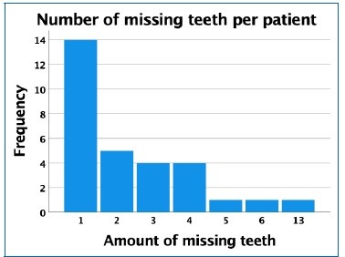
This study obtained a prevalence for tooth agenesis of 20.83%. When excluding the third molars, this value becomes lower, at 11.81%. The most common agenesis corresponded to the maxillary right third molar (tooth 18, absent in 11 patients), followed by tooth 38 (absent in eight patients), and then tooth 35 (absent in seven patients). Figure 1 shows the frequency of the variable “Number of missing teeth per patient.”
It is noteworthy that 60% (18 out of 30) of the participants diagnosed with tooth agenesis in this investigation were male, and males presented a higher prevalence of these anomalies (around 26.1%) than females (16%). No evidence was found indicating that tooth agenesis and sex were not independente (p=0.136).
Regarding the dental arch, 52.63% of all cases of tooth agenesis were located on the mandible. When excluding the third molars, this percentage raised to 57.45%. Given that the expected proportions for tooth agenesis in both arches (if the null hypothesis is accepted) would be 50%, no differences were found for this variable, either including (p=0.646) or excluding the third molars (p=0.307).
Two participants presented supernumerary teeth, resulting in a prevalence of 1.39%. Both cases occurred in the permanente dentition and were found in the same oral cavity region, between the maxillary left lateral incisor and the maxillary left canine. The patients were 7 and 15 years old at the time of their respective radiographs. Neither patient was missing any teeth, and both were male.
Even though this study only found supernumerary teeth in males, no evidence indicated that hyperdontia and sex were not independent (p=0.228). Because of the low number of cases and the location of both supernumerary teeth in the superior arch, their distribution by dental arch resulted in a constant variable that could not be analyzed by the chi-square goodness of fit test.
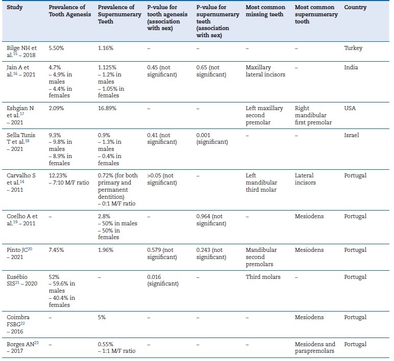
Table 1 presents the results of the literature search performed for comparison with the results of the presente study.1
Discussion
As previously mentioned, tooth agenesis has a reported prevalence of 1.6% to 36.5%.1,2In recent years, most investigations have found a similar value.14-23The values found in this investigation are within the generally accepted ranges for the prevalence of this anomaly.
One participant in this study had 13 missing teeth. The clinical record for this patient showed that genetic studies were being carried out, but no diagnosis was confirmed at the time. Given the association between dental anomalies of number and several genetic entities, it is good practice to investigate the presence of genetic syndromes in cases of oligodontia.2-5,8 If no genetic syndrome is diagnosed, it might also be beneficial to communicate the findings to an oncologist, considering the possibility of an association between oligodontia and some malignant tumors.9-11
Current literature shows that tooth agenesis is more common among female patients.1,2 However, recent studies16,18,21 have found a higher prevalence in males, similar to this investigation. Despite this, our study found no differences to suggest that sex is associated with the diagnosis of tooth agenesis.
This study found that tooth agenesis was more prevalente in the mandibular arch compared to the maxillary arch. This finding agrees with the results reported by Carvalho S, et al.,14 who found a proportion of 14/18 favoring the mandibular arch. The prevalence of 1.39% for the presence of supernumerary teeth in this study fits into the values normally reported for this anomaly - between 0.04% and 3%. Both cases of hyperdontia identified in this investigation were male, and this result agrees with the current literature, which suggests that this anomaly is typically more prevalent in males.3-5No diferences in this study suggested that sex is associated with the diagnosis of supernumerary teeth.
As mentioned earlier, supernumerary teeth are typically classified as mesiodens, paramolars, or distomolars depending on their location,6-8 but the supernumerary teeth found in this investigation do not fit these categories. Still, the results of this study agree with the findings of Carvalho S, et al.,14 who also only reported lateral incisors as supernumerary teeth.
This study was limited by the lack of information in some clinical records, which made it difficult to identify whether some missing teeth were absent due to agenesis or extraction. As a result, there may have been some false positive diagnoses. Besides that, the diagnosis was made exclusively by analyzing orthopantomographs, which are only an auxiliary means of diagnosis. Due to the retrospective nature of this investigation, patients could not be interviewed to determine relevant information regarding their clinical and dental history. These limitations highlight the importance of keeping thorough medical records regarding the patient’s clinical history, as well as the importance of conducting effective interviews for an accurate diagnosis.
The present investigation was able to contribute to the available data regarding the prevalence and characterization of dental anomalies of number in a Portuguese population.
One of the main findings that deviated from the established consensus is the higher prevalence of tooth agenesis among male patients. This finding agrees with some recent studies, as discussed, and may indicate that the previously reported ratios regarding the distribution of this anomaly by sex might be shifting toward being more predominant among males.
However, further research is needed to clarify the potential link between sex and the prevalence of tooth agenesis.
Conclusions
This investigation found a prevalence of 20.83% for tooth agenesis when including third molars and a prevalence of 11.81% when excluding the third molars. Third molars were the teeth most affected by this dental anomaly, followed by the second premolars. Male patients had a higher prevalence of tooth agenesis, and the mandibular arch was the most common location for these anomalies.
The present study also found a prevalence of 1.39% regarding supernumerary teeth. In both cases identified, the only supernumerary tooth in the radiograph was located between the left maxillary lateral incisor and the left maxillary canine. Both patients were male.
The null hypotheses “The variable ‘Diagnosis of Tooth Agenesis’ is independent of the variable ‘Sex,’” “The variable ‘Diagnosis of Supernumerary Teeth’ is independent of the variable ‘Sex,’” and “Tooth agenesis occurs in equal proportions in the maxillary and mandibular arches” were all accepted.

