SciELO - Scientific Electronic Library Online

 
vol.30 número3VExUS na Gestão da Pericardite Constritiva: Relato de CasoProjeto Bem Cuidar no Fim da Vida índice de autoresíndice de assuntosPesquisa de artigos
Home Pagelista alfabética de periódicos  

Serviços Personalizados

Journal

Artigo

Indicadores

Links relacionados

  • Não possue artigos similaresSimilares em SciELO

Compartilhar


Medicina Interna

versão impressa ISSN 0872-671X

Medicina Interna vol.30 no.3 Lisboa set. 2023  Epub 25-Out-2023

https://doi.org/10.24950/rspmi.1703 

Imagens em Medicina/ Images in Medicine

Leishmania in Peripheral Blood: An Unusual and Incidental Find

Leishmania no Sangue Periférico: Um Achado Raro e Incidental

Luis Santos1 
http://orcid.org/0009-0007-3141-4504

Alícia Rodrigues1 

Eliana Silva1 

Vitória Cabral1 
http://orcid.org/0000-0003-1490-7191

1Serviço de Patologia Clínica, Hospital de São José, Centro Hospitalar Universitário Lisboa Central, Lisboa, Portugal


Leishmaniasis is an endemic disease in Portugal, affecting immunocompetent and immunosuppressed patients.1 The gold standard of diagnosis is demonstration of parasites in stained tissues - bone marrow, spleen, lymph nodes, liver, and skin (cutaneous forms) - or in culture. We report a case of accidental discovery of Leishmania in peripheral blood of an immunocompromised patient with several comorbidities.

A 53-years-old man, Caucasian, homeless, with active inhaled drug addiction and a history of heart failure due to the abuse of substances, was admitted into the Department of Infectious Diseases from our Hospitalar Center with a productive cough, dyspnea and diarrhea. Physical examination showed: poor hygiene, emaciated, tachycardia, wheezing at pulmonary auscultation and oral candidiasis; without fever, palpable organomegaly, adenopathy or skin lesions.

Analytically: hemoglobin 99 g/L; white blood cells 4.73 x10⁹/L (neutrophils 76%, lymphocytes 21%); platelets 115 x109/L; Dimer D- 3607 µg/L (RV: < 230); C-reactive protein 31.1 mg/L (RV< 5.0); urine examination positive for cannabinoids, cocaine metabolites and opiates. VIH1 positive: viral charge - 6140000.0 copies/mL, T Helper lymphocytes (CD4+) - 50.1 cells/ µL. Angio-CAT showed ground glass densification areas throughout the lung, suggesting the hypothesis of drug inhalation pneumonitis.

During hospitalization, he was diagnosed with: inaugural HIV/AIDS infection, pneumocystis jiroveci respiratory infection, colitis to Clostridium difficile. He was treated with trimethoprim, sulfamethoxazole, vancomycin, prednisolone and anti-retroviral therapy.

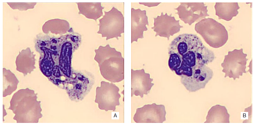
In a routine blood count order, and by observation of the peripheral blood smear, forms of amastigotes of Leishmania spp. were unexpectedly observed (Figs. 1 and 2).

His condition progressed unfavourably with acute hypertensive pulmonary edema, heart failure and progressive renal failure. The patient died on the 21st day of hospitalization.

The appearance of Leishmania in peripheral blood is rare and is indicative of marked and prolonged immunodeficiency, in this case due to HIV infection.

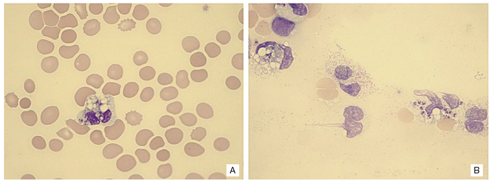
Figure 1: Peripheral blood smears showing amastigotes of Leishmania spp. in the neutrophyls (A) and near to destroided cells (B). May-Grunwald-Giemsa staining, 1000x magnification. 

Figure 2 A and B:  Detail of parasited peripheral blood neutrophils. We can see the oval forms with a dark zone (nucleus) and kinetoplast of the parasites, highly suggestive of Leishmania.2 Coloration: May-Grunwald Giemsa staining 1000x magnification plus zoom (CellaVision). 

REFERENCES

1. Maia C, Campino L. Leishmaniose em Portugal no início do século XXI. An Instit Hig Med Trop. 2018;13:25-8. doi: https://doi.org/10.25761/anai-sihmt.167. [ Links ]

2. Bain B. Blood Cells A Practical Guide. London: Wiley and sons; 2015. [ Links ]

Fontes de Financiamento: Não existiram fontes externas de financiamento para a realização deste artigo.

Confidencialidade dos Dados: Os autores declaram ter seguido os protocolos da sua instituição acerca da publicação dos dados de doentes.

Consentimento: Consentimento do doente para publicação obtido.

Proveniência e Revisão por Pares: Não comissionado; revisão externa por pares.

Financing Support: This work has not received any contribution, grant, or scholarship.

Confidentiality of Data: The authors declare that they have followed the protocols of their work center on the publication of data from patients.

Patient Consent: Consent for publication was obtained.

Provenance and Peer Review: Not commissioned; externally peer re-viewed.

© Autor (es) (ou seu (s) empregador (es)) e Revista SPMI 2023. Reutilização permitida de acordo com CC BY. Nenhuma reutilização comercial. © Author(s) (or their employer(s)) and SPMI Journal 2023. Re-use permitted under CC BY. No commercial re-use.

Received: March 12, 2023; Accepted: April 24, 2023

Correspondence / Correspondência: Vitoria Cabral - vitoria.cabral@netcabo.pt Serviço de Patologia Clínica, Hospital de São José, Centro Hospitalar Universitário Lisboa Central, Lisboa, Portugal Rua José António Serrano, 1150-199 Lisboa

Declaração de Contribuição LS - Pesquisa, redação e aprovação final do artigo. AR - Redação e aprovação final do artigo. ES - Aquisição de imagem, revisão e aprovação final do artigo. VC - Redação, revisão e aprovação final do artigo. Todos os autores aprovaram a versão final a ser publicada. Contributorship Statement LS - Research, writing and final approval of the article. AR - Writing and final approval of the article. ES - Acquisition of image, revision and final approval of the article. VC - Writing, review and final approval of the article. All authors approved the final draft.

Conflitos de Interesse: Os autores declaram a inexistência de conflitos de interesse na realização do presente trabalho.

Conflicts of Interest: The authors have no conflicts of interest to declare.

Creative Commons License This is an open-access article distributed under the terms of the Creative Commons Attribution License