SciELO - Scientific Electronic Library Online

 
vol.32 número2Nódulos eritematosos num recém-nascido: Desafios em contextos particulares índice de autoresíndice de assuntosPesquisa de artigos
Home Pagelista alfabética de periódicos  

Serviços Personalizados

Journal

Artigo

Indicadores

Links relacionados

  • Não possue artigos similaresSimilares em SciELO

Compartilhar


Nascer e Crescer

versão impressa ISSN 0872-0754versão On-line ISSN 2183-9417

Nascer e Crescer vol.32 no.2 Porto jun. 2023  Epub 30-Jun-2023

https://doi.org/10.25753/birthgrowthmj.v32.i2.24637 

Imaging Cases

Imaging clinical case

Caso clínico imagiológico

Diana Henriques Pinto1 
http://orcid.org/0000-0002-8378-5266

Renato Carlos do Vale Ramos2 
http://orcid.org/0009-0005-5604-6213

Mafalda Santos3 
http://orcid.org/0009-0000-5272-9002

1. Department of Pediatrics, Centro Hospitalar Entre Douro e Vouga. 4520-211 Santa Maria da Feira, Portugal. pinto.dianahenriques@gmail.com

2. Department of Orthopedics, Unidade 2, Centro Hospitalar Vila Nova de Gaia/Espinho. 4430-999 Vila Nova de Gaia, Portugal. renato.c.v.ramos@gmail.com

3. Department of Pediatric Orthopedics, Unidade 2, Centro Hospitalar Vila Nova de Gaia/Espinho. 4430-999 Vila Nova de Gaia, Portugal. mafalda.orto@gmail.com


Abstract

A nine-year-old girl with obesity and subclinical hypothyroidism presented with pain in the right thigh with inability to bear weight and associated lameness. These complaints had been present for one month but had recently worsened. There was no history of trauma, fever, or recent infections.

On objective examination, she presented with pain on mobilization of the right thigh that worsened with hip rotation and flexion. Bilateral hip radiographs were obtained and showed right-sided femoral offset between the epiphysis and the neck. These findings were consistent with the diagnosis of slipped capital femoral epiphysis.

The patient was managed with in situ fixation and prophylactic fixation of the contralateral hip, with gradual improvement.

Keywords: hip; obesity; pre-adolescent; slipped capital femoral epiphysis; thigh pain

Resumo

Uma pré-adolescente de nove anos de idade com obesidade e hipotiroidismo subclínico foi observada no Serviço de Urgência por queixas álgicas na coxa direita com um mês de evolução, sem história de trauma, febre ou intercorrências infeciosas.

Ao exame físico, apresentava dor à mobilização da anca direita, sobretudo com rotaçãos e flexão máxima. Foi realizada uma radiografia bilateral da anca, que revelou um deslizamento da epífise proximal do fémur direito em relação ao colo. Estes achados foram consistentes com o diagnóstico dea epifisiólise femoral proximal.

A doente foi tratada com fixação in situ e fixação profilática da anca contralateral, com melhoria progressiva.

Palavras-chave: anca; dor membro inferior; epifisiólise femoral proximal; obesidade; pré-adolescente

A nine-year-old girl presented to the Emergency Department with a 1-month history of limping and thigh pain that had recently worsened. There was no history of trauma or recent infections. The girl had a past medical history of obesity and subclinical hypothyroidism.

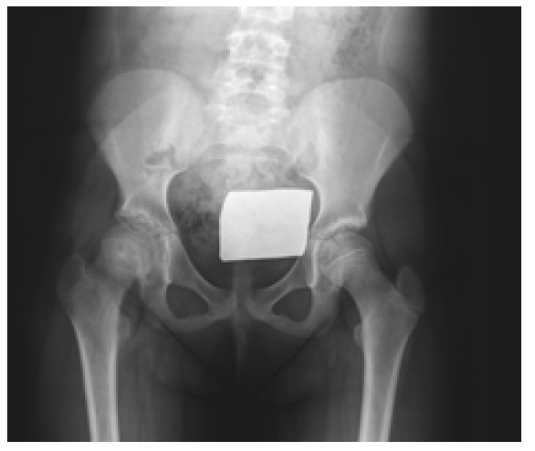
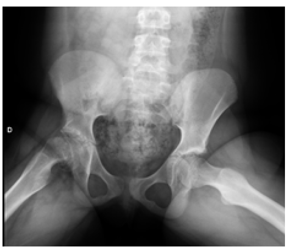
On objective examination, no fever or other systemic signs of infection were observed. The girl had increasing pain with right hip rotation and flection, and therefore bilateral hip radiographs with anteroposterior and lateral frog-leg views were performed (Figures 1and2).

What is your diagnosis?

Figure 1 Anteroposterior view showing height reduction of the right femoral epiphysis 

Figure 2 Frog-leg view showing right femoral offset between epiphysis and neck 

Diagnosis

Slipped capital femoral epiphysis

Discussion

Slipped capital femoral epiphysis (SCFE) is a condition of the proximal femoral physis that results in a slippage of the epiphysis relative to the metaphysis.1,2 It is also called slipped upper femoral epiphysis (SUFE) and is the most common hip disorder in preadolescents and adolescents (between 8 and 15 years of age).1-3 However, it is not commonly observed in routine clinical practice and the diagnosis can be challenging in early stages of disease.1,4

The incidence of SCFE is approximately 10/100,000, ranging from 0,33/100,000 to 24,8/100,000 depending on sex and ethnicity.1,2

The single most significant risk factor for SCFE is obesity (present in 51-77% of cases). Other risk factors include male sex, periods of rapid growth, and prior radiation therapy.5 The average age of onset is 11,2 years in females and 12,0 years in males.6

Ann association has been reported between the development of SCFE and endocrine disorders (including hypothyroidism, hyperthyroidism, hypopituitarism, and growth hormone deficiency) and renal disorders.3,5,6

Approximately 25% of SCFE cases are bilateral, but when unilateral, the left hip is more commonly affected.5,6 Most cases are idiopathic 3,5,6 and often there is no history of trauma or injury prior to symptom onset.3,5,6

The exact underlying mechanism remains uncertain, but it is hypothesized that displacement occurs due to a high physiologic axial load transmitted across a relatively weak physis.6 Conditions such as obesity increase the mechanical weight and force transmitted, while certain endocrine or renal disorders may contribute to weakening of the physis.6

The diagnosis of SCFE is based on symptoms and imaging findings. The most common symptoms are limping and pain that is poorly localized to the hip (52%), thigh (35%), knee (26%), or groin (13,9%).1,3 Distal thigh or knee pain may additionally occur, but the condition most commonly presents as an isolated symptom (15% of cases), which often delays the diagnosis.1

Restricted range of motion of the hip joint is a relevant finding on physical examination, and flexion and medial rotation are often limited. A well-known diagnostic finding is Drehmann's sign, which refers to abduction and lateral rotation of the hip joint in response to flexion.1

To avoid missing the diagnosis of SCFE, a thorough physical examination in the supine position should always be performed on patients who complain of symptoms around the hip joint, thigh, or knee joint. In addition, the mobility of the hip and knee joints should be compared bilaterally.1

The diagnosis is confirmed by bilateral hip radiography, which must include anteroposterior and frog leg lateral views.3

After the diagnosis is established, the correct classification of the disease is required1. SCPE is classified as stable or unstable based on the patient’s ability to bear weight.1-3,9 Classification is very important for prognosis and there is a significant relationship between the delay in diagnosis and an increased severity of dislocation1. The delay in diagnosis is associated with an increased severity of physeal slippage at presentation, and thus poorer long-term outcomes.1,8

The goals of treatment are to prevent slippage progression and complications such as avascular necrosis and chondrolysis.3,4

Stable SCPE is usually treated with in situ screw fixation. Treatment of unstable SCPE usually involves in situ fixation, but there is controversy about the timing of surgery, the need for reduction, and the use of traction.3 Open epiphyseal reduction and fixation may also be performed in these cases.3

In the present case, a prepubertal girl with obesity and subclinical hypothyroidism presented with limping and thigh pain of gradual onset that aggravated with hip rotation and flexion, without history of trauma. This suggested the diagnosis of SCPE, which was subsequently confirmed by radiographic examination. In situ fixation of the affected slippage was performed, as well as prophylactic fixation of the contralateral hip. The patient had a good recovery, with gradual improvement.

Authorship

Diana Henriques Pinto - Conceptualization; Writing - original draft; Writing - review & editing

Renato Carlos do Vale Ramos - Conceptualization; Writing - original draft

Mafalda Santos - Conceptualization; Writing - original draft

References

1. Otani T, Kawaguchi Y, Marumo K. Diagnosis and treatment of slipped capital femoral epiphysis: Recent trends to note. Journal of Orthopaedic Science. 2018;23:220-8. [ Links ]

2. Aprato A, Conti A, Bertolo F, Massè A. Slipped capital femoral epiphysis: current management strategies. Orthopedic Research and Reviews. 2019;11:47-54. [ Links ]

3. Peck D. Slipped Capital Femoral Epiphysis: Diagnosis and Management. American Family Physician. 2010 Aug 1;82(3):258-262. [ Links ]

4. Uvodich M, Schwend R, Stevanovic O, Wurster W, Leamon J, Hermanson A. Patterns of Pain in Adolescents with Slipped Capital Femoral Epiphysis. The Journal of Pediatrics. 2019 Mar;206:184-9. [ Links ]

5. Martinez-Álvarez S, Martínez-González C, Gorozarri C, Abril J, Epeldegui T. Epifisiolisis de la cabeza femoral. Rev Esp Cir Ortop Traumatol. 2012;56(6):506-14. [ Links ]

6. Johns K, Tavarez M. Slipped Capital Femoral Epiphysis. 2020. Available from: https://www.ncbi.nlm.nih.gov/books/NBK538302. [ Links ]

7. Rahme D, Comley A, Foster B, Cindy P. Consequences of diagnostic delays in slipped capital femoral epiphysis. Journal of Pediatric Orthopaedics B. 2006;15:93-7. [ Links ]

8. Barros C, Pinheiro R, Ribeiro E, Duarte R, Varanda P, Maia R. Epifisiólise femoral proximal - Estudo retrospectivo de resultados a longo prazo após fixação in situ. Rev Port Ortop Traum. 2019;27(2):70-8. [ Links ]

9. Loder R, Richards B, Shapiro P, Reznick L, Aronson D. Acute slipped capital femoral epiphysis: the importance of physeal stability. J Bone Joint Surg Am. 1993;75-A:1134-40. [ Links ]

Received: May 26, 2021; Accepted: December 28, 2021

Correspondence to Diana Henriques Pinto Department of Pediatrics Centro Hospitalar Entre Douro e Vouga Rua Dr. Cândido Pinho 5 4520-211 Santa Maria da Feira Email: pinto.dianahenriques@gmail.com

Creative Commons License This is an open-access article distributed under the terms of the Creative Commons Attribution License