


 English (pdf)
 Article in xml format
 Article references
 Send this article by e-mail
 Cited by SciELO
  Similars in
SciELO
Similars in
SciELO 
 Permalink
A female newborn was delivered by forceps at 41 weeks of gestation after an uneventful pregnancy. Birth weight was 3,995 g and Apgar scores were 5/8/10, requiring resuscitation with positive pressure ventilation. The newborn was discharged on the second day after birth.
On the eighth day of life, she was observed in the Pediatric Emergency Department due to a mobile and painless oval paravertebral swelling in the dorsal region with two days of evolution, 20x15 mm, and elastic consistency (Figure I). The remainder physical examination was normal, without rashes or other palpable swelling, and history of fever or lethargy was excluded. Soft-tissue ultrasound was performed, identifying a 13x6x27mm oval hyperechogenic mass in the deep subcutaneous tissue (Figure II). Initial analytical study including blood count and biochemistry was normal.
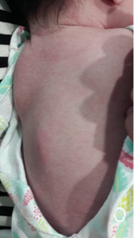
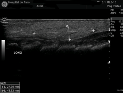
Figure II Hyperechoic oval image of spinous processes with 27x6mm of longitudinal section, visible on dorsal soft tissue ultrasound
Discussion
Subcutaneous fat necrosis is a panniculitis usually arising in the first six weeks of life in healthy term or post-term newborns.1-3 It is characterized by development of single or multiple well-defined nodular lesions of hard elastic consistency, mobile, usually with non-painful borders and sometimes associated with altered skin color.1-4 They are usually located on bony prominences, such as the dorso-lumbar region or upper limbs, and rarely on the buttocks, thighs, and face.1,3,4 Etiology is unknown, although multiple maternal and neonatal risk factors are acknowledged, including pre-eclampsia, gestational diabetes, intra-uterine asphyxia, traumatic delivery, and meconium aspiration.3-7 Two other entities involving subcutaneous fat should be considered in the differential diagnosis, despite being more common in preterm infants. Sclerema neonatorum is a rare panniculitis affecting critically ill preterm and low birthweight newborns, in which the affected skin is tense, mottled purplish, and hard. Another rare condition developing in newborns exposed to cold environments or acute infections during the first week of life is known as scleredema and is characterized by generalized firm, pitting edema preferentially affecting the legs.8
Although subcutaneous fat necrosis is an occasional and self-limited condition with favorable prognosis, it may be associated with severe metabolic complications. Hypercalcemia is the most frequent, occurring in approximately 25% of cases and associated with significant morbidity and mortality.2,5,9 Diagnosis is essentially clinical and follow-up should be directed towards surveillance of potential complications.3-6
Take-home messages
- Subcutaneous fat necrosis is a rare, usually benign, self-limited entity affecting healthy term or post-term neonates.
- Diagnosis is essentially clinical, through recognition of lesion characteristics and risk factors.
- Although unusual, potential complications should be closely monitored and treated, as they can be fatal.

