


 Inglês (pdf)
 Artigo em XML
 Referências do artigo
 Enviar este artigo por email
 Citado por SciELO
  Similares em
SciELO
Similares em
SciELO 
 Permalink
Introduction
Tuberculosis (TB) is a chronic infectious disease caused by various strains of mycobacteria, usually Mycobacterium tuberculosis.1,2 Transmission occurs through airborne infectious particles generated by an infected individual and inhaled by a susceptible individual who may become infected and potentially develop the disease.1,3 Predisposing factors to the disease include crowded urban living, poverty, poor hygiene and health, drug abuse, and immunosuppression, especially by HIV.3,4 Adalimumab, a monoclonal antibody that inhibits the tumor necrosis factor-alpha (TNFα), is also associated with active TB.5
Oral TB is rare and may be either primary or, more often, secondary to pulmonary disease.2,5,6 The disease can manifest in diverse sites in the oral and maxillofacial regions, with notable occurrences observed in the tongue and salivary glands.3
Typically, oral TB presents as an irregular, painful ulcer, often resembling traumatic injury or malignancy, particularly as it gradually enlarges.2,3,7 Despite its infrequency, it must be considered in the differential diagnosis of persistent oral ulcers because tuberculous lesions might be the sole indication of underlying primary or secondary disease.6,8
The treatment of oral tuberculosis lesions is the same as systemic tuberculosis and includes antibiotics such as isoniazid, rifampicin, pyrazinamide, and ethambutol for six months.4 The objective of this study was to report a case of tongue tuberculosis in a 50-year-old male who used adalimumab for five years to treat psoriatic arthritis.
Case report
A 50-year-old man presented in a stomatologist’s office with painful ulcers in his tongue after one year and two months of evolution. Anamnesis revealed a history of psoriatic arthritis treated with adalimumab for five years. Two previous biopsies performed by another professional with a four-month interval did not demonstrate carcinoma but were otherwise inconclusive. The patient was diagnosed with glossitis caused by adalimumab.
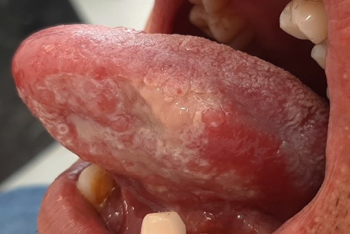
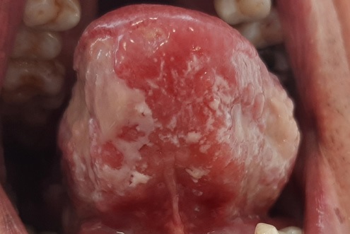
However, four months after discontinuing the medication, the lesions persisted. A new biopsy was scheduled, but the patient refused and sought a second opinion. In the anamnesis, the patient reported no known drug or food allergies and mentioned having frequent unprotected sexual intercourse with different women. There were no systemic signs or symptoms except some weight loss in the past months that, according to him, resulted from his difficulty in eating solid foods due to the intense pain the lesion caused. Vital signs were within the normal range. The extraoral examination revealed a well-nourished man with no evidence of submental, submandibular, or cervical lymphadenopathy. Intraoral examination revealed extensive ulcers, with irregular and indurated borders, situated slightly below the level of the surrounding mucosa in part of the tongue’s dorsum, both borders, and extending to its ventral surface (Figures 1, 2, 3).
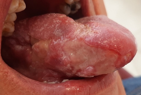
Figure 1 Irregular ulcers on the right border of the tongue extending to its dorsum and ventral region.
Based on the lesion’s clinical presentation and location, the following differential diagnoses were considered: pemphigus vulgaris, oral lichen planus, paracccidioidomycosis, eosinophilic ulcer, oral tuberculosis, and tertiary syphilis. Hematological blood tests (hemogram, FTA-ABS for syphilis, and CA-p24 ELISA for HIV) and sputum culture were requested, and the patient was informed that a new biopsy would be necessary if the exams were all negative.
The patient returned ten days later, and the diagnosis of TB was confirmed through a molecular rapid test (sputum). He was then referred to an infectious disease specialist to initiate treatment comprising isoniazid, rifampicin, pyrazinamide, and ethambutol over six months. In his first consultation, the infectologist requested chest radiography, which revealed no pulmonary involvement. Thus, the diagnosis of primary oral TB was confirmed.
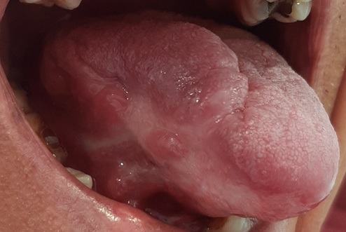
When the patient returned to the dental office after two months of anti-tuberculosis drug therapy, complete regression of tongue ulcers was observed, alongside weight recovery during the treatment period (Figures 4 and 5). After six months, no signs of relapse were observed

Discussion and conclusions
Oral TB is uncommon, accounting for 0.05-1.5% of all TB infections.6 The oral lesions are usually secondarily inoculated with infected sputum or due to hematogenous spread.2,4,6 Primary oral TB without pulmonary involvement, as observed in our patient, is extremely rare and usually poses a diagnostic challenge for the clinician because the oral lesion is the sole manifestation of the disease.2 The tongue is the most commonly affected site, and a non-healing painful ulcer, as observed in the present case, is its main clinical presentation.3,6,7
Accurate diagnosis of TB depends on the detection of Mycobacterium tuberculosis. Clinical and immunological examination, microscopy, radiography, and bacterial cultures are commonly used to screen and diagnose TB.9 A recent systematic review of oral and maxillofacial TB found the diagnostic methods sputum test (26.8%), culture (24.7%), purified protein derivative (PPD), Mantoux test (24.7%), Ziehl-Neelsen (ZN) special staining technique (21.7%), reverse-transcriptase polymerase chain reaction (7.6%), enzyme-linked immunosorbent assay (2%), DNA amplifying for the IS-6110 sequence (1%), Fite Faraco technique (1%), and fluorescence microscopy (0.5%). Of the 301 cases analyzed, only 94 (31.2%) followed the current World Health Organization (WHO) diagnostic recommendations for TB-sputum testing, smear microscopy, culture, or WHO-recommended rapid diagnostic tests, such as the Xpert MTB/RIF assay.3 Sputum culture stands out as a pivotal diagnostic tool for identifying these mycobacteria, boosting a specificity of up to 99% in diagnosing TB. Consequently, it is regarded as the gold standard method among various diagnostic techniques.9
Psoriasis, a prevalent immune-mediated condition characterized by chronicity, treatment resistance, and systemic involvement, significantly impacts patients’ quality of life.10 Over recent years, adalimumab has emerged as a potent therapy for individuals with psoriatic arthritis.11 The therapeutic action of adalimumab and other TNFα inhibitors may influence T cell dynamics, function, and cytokine signaling, which are crucial for controlling TB infections or sustaining granulomas.12 Such modulation can potentially predispose patients to latent TB reactivation or new infections, particularly notable in regions with low TB prevalence.10 This scenario was exemplified in our patient residing in a low TB risk area in Southern Brazil, who had been on adalimumab therapy for five years.
Due to this high rate of TB conversion among psoriasis patients, some authors recommend TB reevaluation after the first 3 months and then semiannually for the following 2 years in patients under adalimumab.10 Our patient reported no monitoring for the disease.
Despite the absence of systemic illness and its rareness, oral TB should always be considered in the differential diagnosis of persistent oral ulcers. Dentists should be aware that adalimumab increases the risk of TB reactivation or acquisition of new TB disease and pay special attention to patients under this medication. Early diagnosis is fundamental to reduce the spread of infection in the community and avoid systemic complications, given that the disease is still a leading cause of death among infectious diseases.

